1STC

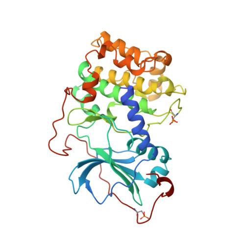
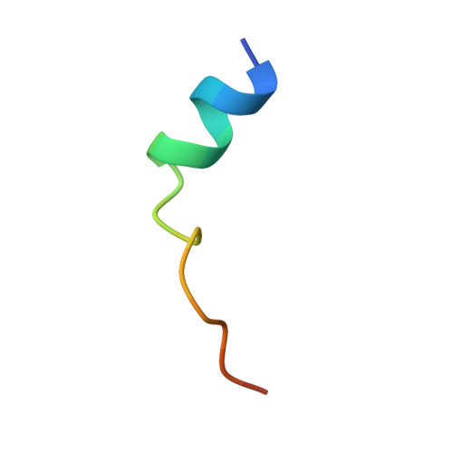
CAMP-DEPENDENT PROTEIN KINASE, ALPHA-CATALYTIC SUBUNIT IN COMPLEX WITH STAUROSPORINE


Experimental Data Snapshot

  • Method: X-RAY DIFFRACTION
  • Resolution: 2.30 Å
  • R-Value Free: 0.330 
  • R-Value Work: 0.208 
  • R-Value Observed: 0.208 

Starting Model: experimental
View more details

wwPDB Validation   3D Report Full Report


This is version 1.4 of the entry. See complete history


Literature

Staurosporine-induced conformational changes of cAMP-dependent protein kinase catalytic subunit explain inhibitory potential.

Prade, L.Engh, R.A.Girod, A.Kinzel, V.Huber, R.Bossemeyer, D.

(1997) Structure 5: 1627-1637

  • DOI: https://doi.org/10.1016/s0969-2126(97)00310-9
  • Primary Citation of Related Structures:  
    1STC

  • PubMed Abstract: 

    Staurosporine inhibits most protein kinases at low nanomolar concentrations. As most tyrosine kinases, along with many serine/threonine kinases, are either proto oncoproteins or are involved in oncogenic signaling, the development of protein kinase inhibitors is a primary goal of cancer research. Staurosporine and many of its derivatives have significant biological effects, and are being tested as anticancer drugs. To understand in atomic detail the mode of inhibition and the parameters of high-affinity binding of staurosporine to protein kinases, the molecule was cocrystallized with the catalytic subunit of cAMP-dependent protein kinase. The crystal structure of the protein kinase catalytic subunit with staurosporine bound to the adenosine pocket shows considerable induced-fit rearrangement of the enzyme and a unique open conformation. The inhibitor mimics several aspects of adenosine binding, including both polar and nonpolar interactions with enzyme residues, and induces conformational changes of neighboring enzyme residues. The results explain the high inhibitory potency of staurosporine, and also illustrate the flexibility of the protein kinase active site. The structure, therefore, is not only useful for the design of improved anticancer therapeutics and signaling drugs, but also provides a deeper understanding of the conformational flexibility of the protein kinase.


  • Organizational Affiliation

    Abteilung Strukturforschung Max-Planck-Institut für Biochemie, Martinsried, Germany.


Macromolecules
Find similar proteins by:  (by identity cutoff)  |  3D Structure
Entity ID: 1
MoleculeChains Sequence LengthOrganismDetailsImage
CAMP-DEPENDENT PROTEIN KINASEA [auth E]350Bos taurusMutation(s): 2 
EC: 2.7.1.37 (PDB Primary Data), 2.7.11.11 (UniProt)
UniProt
Find proteins for P00517 (Bos taurus)
Explore P00517 
Go to UniProtKB:  P00517
Entity Groups  
Sequence Clusters30% Identity50% Identity70% Identity90% Identity95% Identity100% Identity
UniProt GroupP00517
Sequence Annotations
Expand
  • Reference Sequence

Find similar proteins by:  Sequence   |   3D Structure  

Entity ID: 2
MoleculeChains Sequence LengthOrganismDetailsImage
PROTEIN KINASE INHIBITORB [auth I]20Oryctolagus cuniculusMutation(s): 0 
Gene Names: PKIA
UniProt
Find proteins for P61926 (Oryctolagus cuniculus)
Explore P61926 
Go to UniProtKB:  P61926
Entity Groups  
Sequence Clusters30% Identity50% Identity70% Identity90% Identity95% Identity100% Identity
UniProt GroupP61926
Sequence Annotations
Expand
  • Reference Sequence
Small Molecules
Ligands 1 Unique
IDChains Name / Formula / InChI Key2D Diagram3D Interactions
STU
Query on STU

Download Ideal Coordinates CCD File 
C [auth E]STAUROSPORINE
C28 H26 N4 O3
HKSZLNNOFSGOKW-FYTWVXJKSA-N
Modified Residues  2 Unique
IDChains TypeFormula2D DiagramParent
SEP
Query on SEP
A [auth E]L-PEPTIDE LINKINGC3 H8 N O6 PSER
TPO
Query on TPO
A [auth E]L-PEPTIDE LINKINGC4 H10 N O6 PTHR
Experimental Data & Validation

Experimental Data

  • Method: X-RAY DIFFRACTION
  • Resolution: 2.30 Å
  • R-Value Free: 0.330 
  • R-Value Work: 0.208 
  • R-Value Observed: 0.208 
  • Space Group: P 21 21 21
Unit Cell:
Length ( Å )Angle ( ˚ )
a = 68.99α = 90
b = 72.82β = 90
c = 76.63γ = 90
Software Package:
Software NamePurpose
X-PLORmodel building
X-PLORrefinement
MOSFLMdata reduction
Agrovatadata scaling
ROTAVATAdata scaling
X-PLORphasing

Structure Validation

View Full Validation Report


Entry History 

Deposition Data

Revision History  (Full details and data files)

  • Version 1.0: 1998-02-25
    Type: Initial release
  • Version 1.1: 2008-03-03
    Changes: Version format compliance
  • Version 1.2: 2011-07-13
    Changes: Version format compliance
  • Version 1.3: 2023-08-09
    Changes: Database references, Derived calculations, Other, Refinement description
  • Version 1.4: 2024-11-20
    Changes: Data collection, Structure summary