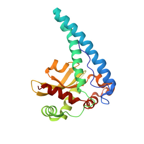
Role of hydrogen bonding in the active site of human manganese superoxide dismutase.
Greenleaf, W.B., Perry, J.J., Hearn, A.S., Cabelli, D.E., Lepock, J.R., Stroupe, M.E., Tainer, J.A., Nick, H.S., Silverman, D.N.(2004) Biochemistry 43: 7038-7045
- PubMed: 15170341
- DOI: https://doi.org/10.1021/bi049888k
- Primary Citation of Related Structures:
1SZX - PubMed Abstract:
The side chain of Gln143, a conserved residue in manganese superoxide dismutase (MnSOD), forms a hydrogen bond with the manganese-bound solvent and is critical in maintaining catalytic activity. The side chains of Tyr34 and Trp123 form hydrogen bonds with the carboxamide of Gln143. We have replaced Tyr34 and Trp123 with Phe in single and double mutants of human MnSOD and measured their catalytic activity by stopped-flow spectrophotometry and pulse radiolysis. The replacements of these side chains inhibited steps in the catalysis as much as 50-fold; in addition, they altered the gating between catalysis and formation of a peroxide complex to yield a more product-inhibited enzyme. The replacement of both Tyr34 and Trp123 in a double mutant showed that these two residues interact cooperatively in maintaining catalytic activity. The crystal structure of Y34F/W123F human MnSOD at 1.95 A resolution suggests that this effect is not related to a conformational change in the side chain of Gln143, which does not change orientation in Y34F/W123F, but rather to more subtle electronic effects due to the loss of hydrogen bonding to the carboxamide side chain of Gln143. Wild-type MnSOD containing Trp123 and Tyr34 has approximately the same thermal stability compared with mutants containing Phe at these positions, suggesting the hydrogen bonds formed by these residues have functional rather than structural roles.
- Departments of Pharmacology and Neuroscience, University of Florida, Gainesville, Florida 32610-0267, USA.
Organizational Affiliation:


