1T18

Early intermediate IE1 from time-resolved crystallography of the E46Q mutant of PYP


Experimental Data Snapshot

  • Method: X-RAY DIFFRACTION
  • Resolution: 1.60 Å
  • R-Value Free: 
    0.247 (Depositor), 0.240 (DCC) 
  • R-Value Work: 
    0.227 (Depositor), 0.230 (DCC) 
  • R-Value Observed: 
    0.229 (Depositor) 

wwPDB Validation   3D Report Full Report


Ligand Structure Quality Assessment 

Created with Raphaël 2.3.0Worse 01 BetterLigand structure goodness of fit to experimental dataBest fitted HC4Click on this verticalbar to view details

This is version 1.5 of the entry. See complete history


Literature

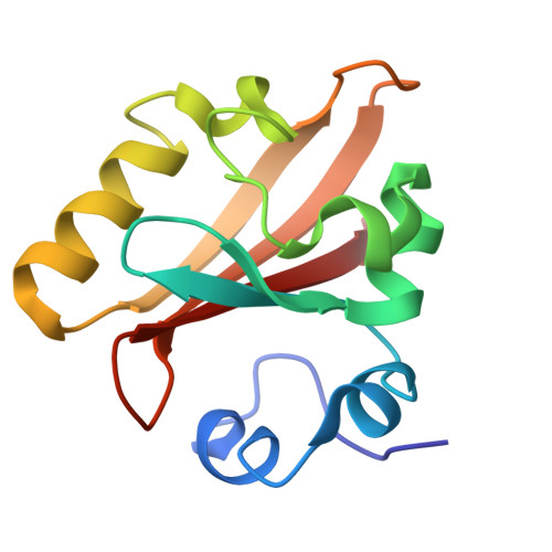
A Structural Pathway for Signaling in the E46Q Mutant of Photoactive Yellow Protein

Rajagopal, S.Anderson, S.Srajer, V.Schmidt, M.Pahl, R.Moffat, K.

(2005) Structure 13: 55-63

  • DOI: https://doi.org/10.1016/j.str.2004.10.016
  • Primary Citation of Related Structures:  
    1T18, 1T19, 1T1A, 1T1B, 1T1C

  • PubMed Abstract: 

    In the bacterial photoreceptor photoactive yellow protein (PYP), absorption of blue light by its chromophore leads to a conformational change in the protein associated with differential signaling activity, as it executes a reversible photocycle. Time-resolved Laue crystallography allows structural snapshots (as short as 150 ps) of high crystallographic resolution (approximately 1.6 A) to be taken of a protein as it functions. Here, we analyze by singular value decomposition a comprehensive time-resolved crystallographic data set of the E46Q mutant of PYP throughout the photocycle spanning 10 ns-100 ms. We identify and refine the structures of five distinct intermediates and provide a plausible chemical kinetic mechanism for their inter conversion. A clear structural progression is visible in these intermediates, in which a signal generated at the chromophore propagates through a distinct structural pathway of conserved residues and results in structural changes near the N terminus, over 20 A distant from the chromophore.


  • Organizational Affiliation

    Department of Biochemistry and Molecular Biology, University of Chicago, Illinois 60637, USA.


Macromolecules
Find similar proteins by:  (by identity cutoff)  |  3D Structure
Entity ID: 1
MoleculeChains Sequence LengthOrganismDetailsImage
Photoactive yellow protein125Halorhodospira halophilaMutation(s): 1 
Gene Names: PYP
UniProt
Find proteins for P16113 (Halorhodospira halophila)
Explore P16113 
Go to UniProtKB:  P16113
Entity Groups  
Sequence Clusters30% Identity50% Identity70% Identity90% Identity95% Identity100% Identity
UniProt GroupP16113
Sequence Annotations
Expand
  • Reference Sequence
Small Molecules
Ligands 1 Unique
IDChains Name / Formula / InChI Key2D Diagram3D Interactions
HC4
Query on HC4

Download Ideal Coordinates CCD File 
B [auth A]4'-HYDROXYCINNAMIC ACID
C9 H8 O3
NGSWKAQJJWESNS-ZZXKWVIFSA-N
Experimental Data & Validation

Experimental Data

  • Method: X-RAY DIFFRACTION
  • Resolution: 1.60 Å
  • R-Value Free:  0.247 (Depositor), 0.240 (DCC) 
  • R-Value Work:  0.227 (Depositor), 0.230 (DCC) 
  • R-Value Observed: 0.229 (Depositor) 
Space Group: P 63
Unit Cell:
Length ( Å )Angle ( ˚ )
a = 66.917α = 90
b = 66.917β = 90
c = 41.002γ = 120
Software Package:
Software NamePurpose
SHELXmodel building
SHELXL-97refinement
LaueViewdata scaling

Structure Validation

View Full Validation Report


Ligand Structure Quality Assessment 

Created with Raphaël 2.3.0Worse 01 BetterLigand structure goodness of fit to experimental dataBest fitted HC4Click on this verticalbar to view details

Entry History 

Deposition Data

Revision History  (Full details and data files)

  • Version 1.0: 2005-01-18
    Type: Initial release
  • Version 1.1: 2008-04-30
    Changes: Version format compliance
  • Version 1.2: 2011-07-13
    Changes: Version format compliance
  • Version 1.3: 2015-09-09
    Changes: Other
  • Version 1.4: 2017-10-11
    Changes: Refinement description
  • Version 1.5: 2021-10-27
    Changes: Database references, Derived calculations