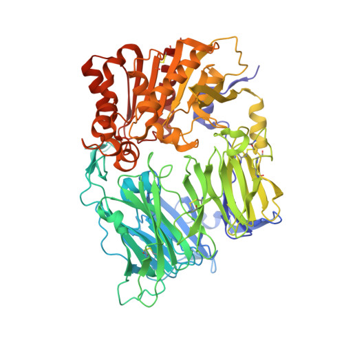
1TK3

Crystal Structure Of Human Apo Dipeptidyl Peptidase IV/CD26


Experimental Data Snapshot

  • Method: X-RAY DIFFRACTION
  • Resolution: 2.00 Å
  • R-Value Free: 0.272 
  • R-Value Work: 0.229 
  • R-Value Observed: 0.229 

Starting Model: experimental
View more details

wwPDB Validation   3D Report Full Report


Ligand Structure Quality Assessment 


This is version 2.1 of the entry. See complete history


Literature

Tyrosine 547 Constitutes an Essential Part of the Catalytic Mechanism of Dipeptidyl Peptidase IV

Bjelke, J.R.Christensen, J.Branner, S.Wagtmann, N.Olsen, C.Kanstrup, A.B.Rasmussen, H.B.

(2004) J Biol Chem 279: 34691-34697

  • DOI: https://doi.org/10.1074/jbc.M405400200
  • Primary Citation of Related Structures:  
    1TK3, 1TKR, 1U8E

  • PubMed Abstract: 

    Human dipeptidyl peptidase IV (DPP-IV) is a ubiquitously expressed type II transmembrane serine protease. It cleaves the penultimate positioned prolyl bonds at the N terminus of physiologically important peptides such as the incretin hormones glucagon-like peptide 1 and glucose-dependent insulinotropic peptide. In this study, we have characterized different active site mutants. The Y547F mutant as well as the catalytic triad mutants S630A, D708A, and H740L showed less than 1% wild type activity. X-ray crystal structure analysis of the Y547F mutant revealed no overall changes compared with wild type apoDPP-IV, except the ablation of the hydroxyl group of Tyr(547) and a water molecule positioned in close proximity to Tyr(547). To elucidate further the reaction mechanism, we determined the crystal structure of DPP-IV in complex with diisopropyl fluorophosphate, mimicking the tetrahedral intermediate. The kinetic and structural findings of the tyrosine residue are discussed in relation to the catalytic mechanism of DPP-IV and to the inhibitory mechanism of the 2-cyanopyrrolidine class of potent DPP-IV inhibitors, proposing an explanation for the specificity of this class of inhibitors for the S9b family among serine proteases.


  • Organizational Affiliation

    Institute of Medical Biochemistry and Genetics, Panum Institute, University of Copenhagen, Bledamsvej 3C, DK-2200 Copenhagen, Denmark.


Macromolecules
Find similar proteins by:  (by identity cutoff)  |  3D Structure
Entity ID: 1
MoleculeChains Sequence LengthOrganismDetailsImage
Dipeptidyl peptidase IV
A, B
728Homo sapiensMutation(s): 0 
Gene Names: DPP4ADCP2CD26
EC: 3.4.14.5
UniProt & NIH Common Fund Data Resources
Find proteins for P27487 (Homo sapiens)
Explore P27487 
Go to UniProtKB:  P27487
PHAROS:  P27487
GTEx:  ENSG00000197635 
Entity Groups  
Sequence Clusters30% Identity50% Identity70% Identity90% Identity95% Identity100% Identity
UniProt GroupP27487
Glycosylation
Glycosylation Sites: 7Go to GlyGen: P27487-1
Sequence Annotations
Expand
  • Reference Sequence
Oligosaccharides

Help

Entity ID: 2
MoleculeChains Length2D Diagram Glycosylation3D Interactions
2-acetamido-2-deoxy-beta-D-glucopyranose-(1-4)-[alpha-L-fucopyranose-(1-6)]2-acetamido-2-deoxy-beta-D-glucopyranose
C, D, I
3N-Glycosylation
Glycosylation Resources
GlyTouCan:  G21290RB
GlyCosmos:  G21290RB
GlyGen:  G21290RB
Entity ID: 3
MoleculeChains Length2D Diagram Glycosylation3D Interactions
2-acetamido-2-deoxy-beta-D-glucopyranose-(1-4)-2-acetamido-2-deoxy-beta-D-glucopyranose
E, G, H, J
2N-Glycosylation
Glycosylation Resources
GlyTouCan:  G42666HT
GlyCosmos:  G42666HT
GlyGen:  G42666HT
Entity ID: 4
MoleculeChains Length2D Diagram Glycosylation3D Interactions
alpha-D-mannopyranose-(1-4)-2-acetamido-2-deoxy-alpha-D-glucopyranose-(1-4)-2-acetamido-2-deoxy-beta-D-glucopyranose
F
3N-Glycosylation
Glycosylation Resources
GlyTouCan:  G52260VY
GlyCosmos:  G52260VY
GlyGen:  G52260VY
Entity ID: 5
MoleculeChains Length2D Diagram Glycosylation3D Interactions
beta-D-mannopyranose-(1-4)-2-acetamido-2-deoxy-beta-D-glucopyranose-(1-4)-2-acetamido-2-deoxy-beta-D-glucopyranose
K, L
3N-Glycosylation
Glycosylation Resources
GlyTouCan:  G15407YE
GlyCosmos:  G15407YE
GlyGen:  G15407YE
Entity ID: 6
MoleculeChains Length2D Diagram Glycosylation3D Interactions
2-acetamido-2-deoxy-alpha-D-glucopyranose-(1-4)-2-acetamido-2-deoxy-beta-D-glucopyranose
M
2N-Glycosylation
Glycosylation Resources
GlyTouCan:  G07375KG
GlyCosmos:  G07375KG
GlyGen:  G07375KG
Experimental Data & Validation

Experimental Data

  • Method: X-RAY DIFFRACTION
  • Resolution: 2.00 Å
  • R-Value Free: 0.272 
  • R-Value Work: 0.229 
  • R-Value Observed: 0.229 
  • Space Group: P 21 21 21
Unit Cell:
Length ( Å )Angle ( ˚ )
a = 119.264α = 90
b = 122.36β = 90
c = 129.823γ = 90
Software Package:
Software NamePurpose
HKL-2000data collection
SCALEPACKdata scaling
CNXrefinement
HKL-2000data reduction
CNXphasing

Structure Validation

View Full Validation Report


Ligand Structure Quality Assessment 


Entry History 

Deposition Data

Revision History  (Full details and data files)

  • Version 1.0: 2004-07-06
    Type: Initial release
  • Version 1.1: 2008-04-30
    Changes: Version format compliance
  • Version 1.2: 2011-07-13
    Changes: Non-polymer description, Version format compliance
  • Version 1.3: 2016-11-30
    Changes: Other
  • Version 2.0: 2020-07-29
    Type: Remediation
    Reason: Carbohydrate remediation
    Changes: Advisory, Atomic model, Data collection, Derived calculations, Structure summary
  • Version 2.1: 2023-08-23
    Changes: Data collection, Database references, Refinement description, Structure summary