Superagonistic Action of 14-epi-Analogs of 1,25-Dihydroxyvitamin D Explained by Vitamin D Receptor-Coactivator Interaction
Eelen, G., Verlinden, L., Rochel, N., Claessens, F., De Clercq, P., Vandewalle, M., Tocchini-Valentini, G., Moras, D., Bouillon, R., Verstuyf, A.(2005) Mol Pharmacol 67: 1566-1573
- PubMed: 15728261 Search on PubMed
- DOI: https://doi.org/10.1124/mol.104.008730
- Primary Citation Related Structures:
1TXI - PubMed Abstract:
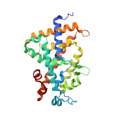
Two 14-epi-analogs of 1,25-dihydroxyvitamin D3 [1,25-(OH)(2)D(3)], 19-nor-14-epi-23-yne-1,25-(OH)2D3 (TX522) and 19-nor-14,20-bisepi-23-yne-1,25-(OH)2D3 (TX527), show enhanced antiproliferative (at least 10-fold) and markedly lower calcemic effects both in vitro and in vivo, compared with 1,25-(OH)2D3. This study aimed to evaluate their superagonistic effect at the level of interaction between the Vitamin D receptor (VDR) and coactivators. Mammalian two-hybrid assays with VP16-fused VDR and GAL4-DNA-binding-domain-fused steroid receptor coactivator 1 (SRC-1), transcriptional intermediary factor 2 (Tif2), or DRIP205 showed the 14-epi-analogs to be more potent inducers of VDR-coactivator interactions than 1,25-(OH)2D3 (up to 16- and 20-fold stronger induction of VDR-SRC-1 interaction for TX522 and TX527 at 10(-10) M). Similar assays in which metabolism of 1,25-(OH)2D3 was blocked with VID400, a selective inhibitor of the 1,25-(OH)2D3-metabolizing enzyme CYP24, showed that the enhanced potency of these analogs in establishing VDR-coactivator interactions can only partially be accounted for by their increased resistance to metabolic degradation. Crystallization of TX522 complexed to the ligand binding domain of the human VDR demonstrated that the epi-configuration of C14 caused the CD ring of the ligand to shift by 0.5 angstroms, thereby bringing the C12 atom into closer contact with Val300. Moreover, C22 of TX522 made an additional contact with the CD1 atom of Ile268 because of the rigidity of the triple bond-containing side chain. The position and conformation of the activation helix H12 of VDR was strictly maintained. In conclusion, this study provides deeper insight into the docking of TX522 in the LBP and shows that stronger VDR-coactivator interactions underlie the superagonistic activity of the two 14-epi-analogs.
- Laboratorium voor Experimentele Geneeskunde en Endocrinologie, Katholieke Universiteit Leuven, Belgium.
Organizational Affiliation: