1U3V | pdb_00001u3v

Crystal Structure of Human Alcohol Dehydrogenase Beta-1-Beta-1 Isoform Complexed with N-Heptylformamide Determined to 1.65 Angstrom Resolution


Experimental Data Snapshot

  • Method: X-RAY DIFFRACTION
  • Resolution: 1.65 Å
  • R-Value Free: 
    0.185 (Depositor), 0.181 (DCC) 
  • R-Value Work: 
    0.164 (Depositor), 0.161 (DCC) 
  • R-Value Observed: 
    0.165 (Depositor) 

Starting Model: experimental
View more details

wwPDB Validation 3D Report Full Report

Validation slider image for 1U3V

Ligand Structure Quality Assessment 


This is version 1.4 of the entry. See complete history

Literature

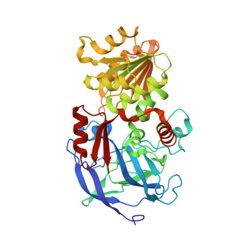
Structure of three class I human alcohol dehydrogenases complexed with isoenzyme specific formamide inhibitors

Gibbons, B.J.Hurley, T.D.

(2004) Biochemistry 43: 12555-12562

  • DOI: https://doi.org/10.1021/bi0489107
  • Primary Citation Related Structures: 
    1U3T, 1U3U, 1U3V, 1U3W

  • PubMed Abstract: 

    Formamides are aldehyde analogues that have demonstrated potent and selective inhibition of human alcohol dehydrogenase isoenzymes. The alphaalpha, beta(1)beta(1), gamma(2)gamma(2), and sigmasigma isoforms have all been found to be strongly inhibited by substituted formamides. In this paper, the structure of the alphaalpha isoform of human alcohol dehydrogenase complexed with N-cyclopentyl-N-cyclobutylformamide was determined by X-ray crystallography to 2.5 A resolution, the beta(1)beta(1) isoform of human alcohol dehydrogenase complexed with N-benzylformamide and with N-heptylformamide was determined to 1.6 and 1.65 A resolution, respectively, and the structure of the gamma(2)gamma(2) isoform complexed with N-1-methylheptylformamide was determined to 1.45 A resolution. These structures provide the first substrate-level view of the local structural differences that give rise to the individual substrate preferences shown by these highly related isoenzymes. Consistent with previous work, the carbonyl oxygen of the inhibitors interacts directly with the catalytic zinc and the hydroxyl group of Thr48 (Ser48 for gamma(2)gamma(2)) of the enzyme. The benzene ring of N-benzylformamide and the carbon chains of N-heptylformamide and N-1-methylheptylformamide interact with the sides of the hydrophobic substrate pocket whose size and shape is dictated by residue exchanges between the beta(1)beta(1) and gamma(2)gamma(2) isoenzymes. In particular, the exchange of Ser for Thr at position 48 and the exchange of Val for Leu at position 141 in the gamma(2)gamma(2) isoenzyme create an environment with stereoselectivity for the R-enantiomer of the branched N-1-methylheptylformamide inhibitor in this isoenzyme. The primary feature of the alphaalpha isoform is the Ala for Phe93 exchange that enlarges the active site near the catalytic zinc and creates the specificity for the branched N-cyclopentyl-N-cyclobutylformamide inhibitor, which shows the greatest selectivity for this unique isoenzyme of any of the formamide inhibitors.


  • Organizational Affiliation
    • Department of Biochemistry and Molecular Biology, Indiana University School of Medicine, 635 Barnhill Drive, Room MS 4017, Indianapolis, Indiana 46202-5122, USA.

Macromolecule Content 

  • Total Structure Weight: 81.52 kDa 
  • Atom Count: 6,731 
  • Modeled Residue Count: 748 
  • Deposited Residue Count: 748 
  • Unique protein chains: 1

Macromolecules

Find similar proteins by:|  3D Structure
Entity ID: 1
MoleculeChains  Sequence LengthOrganismDetailsImage
Alcohol dehydrogenase beta chain
A, B
374Homo sapiensMutation(s): 0 
Gene Names: ADH1B
EC: 1.1.1.1 (PDB Primary Data), 1.1.1.105 (UniProt)
UniProt & NIH Common Fund Data Resources
Find proteins for P00325 (Homo sapiens)
Explore P00325 
Go to UniProtKB:  P00325
PHAROS:  P00325
GTEx:  ENSG00000196616 
Entity Groups
Sequence Clusters30% Identity50% Identity70% Identity90% Identity95% Identity100% Identity
UniProt GroupP00325
Sequence Annotations
Expand
Reference Sequence

Small Molecules

Ligands 4 Unique
IDChains Name / Formula / InChI Key2D Diagram3D Interactions
NAD

Query on NAD


Download:Ideal Coordinates CCD File
E [auth A],
J [auth B]
NICOTINAMIDE-ADENINE-DINUCLEOTIDE
C21 H27 N7 O14 P2
BAWFJGJZGIEFAR-NNYOXOHSSA-N
HPL

Query on HPL


Download:Ideal Coordinates CCD File
F [auth A],
K [auth B]
HEPTYLFORMAMIDE
C8 H17 N O
YAUHDTOEJHVKJO-UHFFFAOYSA-N
PO4

Query on PO4


Download:Ideal Coordinates CCD File
I [auth B]PHOSPHATE ION
O4 P
NBIIXXVUZAFLBC-UHFFFAOYSA-K
ZN

Query on ZN


Download:Ideal Coordinates CCD File
C [auth A],
D [auth A],
G [auth B],
H [auth B]
ZINC ION
Zn
PTFCDOFLOPIGGS-UHFFFAOYSA-N

Experimental Data & Validation

Experimental Data

  • Method: X-RAY DIFFRACTION
  • Resolution: 1.65 Å
  • R-Value Free:  0.185 (Depositor), 0.181 (DCC) 
  • R-Value Work:  0.164 (Depositor), 0.161 (DCC) 
  • R-Value Observed: 0.165 (Depositor) 
Space Group: P 1
Unit Cell:
Length ( Å )Angle ( ˚ )
a = 44.32α = 79.58
b = 52.83β = 89.43
c = 90.35γ = 68.6
Software Package:
Software NamePurpose
DENZOdata reduction
SCALEPACKdata scaling
TRUNCATEdata reduction
AMoREphasing
CNSrefinement
CCP4data scaling

Structure Validation

View Full Validation Report


Ligand Structure Quality Assessment 


Entry History 

Deposition Data

Revision History  (Full details and data files)

  • Version 1.0: 2004-10-26
    Type: Initial release
  • Version 1.1: 2008-04-30
    Changes: Version format compliance
  • Version 1.2: 2011-07-13
    Changes: Version format compliance
  • Version 1.3: 2017-10-11
    Changes: Refinement description
  • Version 1.4: 2023-10-25
    Changes: Data collection, Database references, Derived calculations, Refinement description