1U4H

Crystal structure of an oxygen binding H-NOX domain related to soluble guanylate cyclases (oxygen complex)


Experimental Data Snapshot

  • Method: X-RAY DIFFRACTION
  • Resolution: 2.07 Å
  • R-Value Free: 
    0.255 (Depositor), 0.254 (DCC) 
  • R-Value Work: 
    0.216 (Depositor), 0.216 (DCC) 

wwPDB Validation   3D Report Full Report


Ligand Structure Quality Assessment 

Created with Raphaël 2.3.0Worse 01 BetterLigand structure goodness of fit to experimental dataBest fitted HEMClick on this verticalbar to view details

This is version 1.3 of the entry. See complete history


Literature

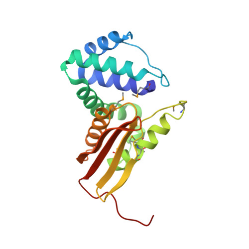
Crystal structure of an oxygen-binding heme domain related to soluble guanylate cyclases.

Pellicena, P.Karow, D.S.Boon, E.M.Marletta, M.A.Kuriyan, J.

(2004) Proc Natl Acad Sci U S A 101: 12854-12859

  • DOI: https://doi.org/10.1073/pnas.0405188101
  • Primary Citation of Related Structures:  
    1U4H, 1U55, 1U56

  • PubMed Abstract: 

    Soluble guanylate cyclases are nitric oxide-responsive signaling proteins in which the nitric oxide sensor is a heme-binding domain of unknown structure that we have termed the heme-NO and oxygen binding (H-NOX) domain. H-NOX domains are also found in bacteria, either as isolated domains, or are fused through a membrane-spanning region to methyl-accepting chemotaxis proteins. We have determined the crystal structure of an oxygen-binding H-NOX domain of one such signaling protein from the obligate anaerobe Thermoanaerobacter tengcongensis at 1.77-angstroms resolution, revealing a protein fold unrelated to known structures. Particularly striking is the structure of the protoporphyrin IX group, which is distorted from planarity to an extent not seen before in protein-bound heme groups. Comparison of the structure of the H-NOX domain in two different crystal forms suggests a mechanism whereby alteration in the degree of distortion of the heme group is coupled to changes on the molecular surface of the H-NOX domain and potentially to changes in intermolecular interactions.


  • Organizational Affiliation

    Department of Molecular and Cell Biology, University of California, Berkeley, CA 94720, USA.


Macromolecules
Find similar proteins by:  (by identity cutoff)  |  3D Structure
Entity ID: 1
MoleculeChains Sequence LengthOrganismDetailsImage
Heme-based Methyl-accepting Chemotaxis Protein
A, B
188Caldanaerobacter subterraneus subsp. tengcongensisMutation(s): 7 
Gene Names: Tar4
UniProt
Find proteins for Q8RBX6 (Caldanaerobacter subterraneus subsp. tengcongensis (strain DSM 15242 / JCM 11007 / NBRC 100824 / MB4))
Explore Q8RBX6 
Go to UniProtKB:  Q8RBX6
Entity Groups  
Sequence Clusters30% Identity50% Identity70% Identity90% Identity95% Identity100% Identity
UniProt GroupQ8RBX6
Sequence Annotations
Expand
  • Reference Sequence
Small Molecules
Ligands 4 Unique
IDChains Name / Formula / InChI Key2D Diagram3D Interactions
HEM
Query on HEM

Download Ideal Coordinates CCD File 
C [auth A],
I [auth B]
PROTOPORPHYRIN IX CONTAINING FE
C34 H32 Fe N4 O4
KABFMIBPWCXCRK-RGGAHWMASA-L
GOL
Query on GOL

Download Ideal Coordinates CCD File 
E [auth A],
F [auth A],
G [auth A],
K [auth B]
GLYCEROL
C3 H8 O3
PEDCQBHIVMGVHV-UHFFFAOYSA-N
SCN
Query on SCN

Download Ideal Coordinates CCD File 
H [auth B]THIOCYANATE ION
C N S
ZMZDMBWJUHKJPS-UHFFFAOYSA-M
OXY
Query on OXY

Download Ideal Coordinates CCD File 
D [auth A],
J [auth B]
OXYGEN MOLECULE
O2
MYMOFIZGZYHOMD-UHFFFAOYSA-N
Modified Residues  1 Unique
IDChains TypeFormula2D DiagramParent
MSE
Query on MSE
A, B
L-PEPTIDE LINKINGC5 H11 N O2 SeMET
Experimental Data & Validation

Experimental Data

  • Method: X-RAY DIFFRACTION
  • Resolution: 2.07 Å
  • R-Value Free:  0.255 (Depositor), 0.254 (DCC) 
  • R-Value Work:  0.216 (Depositor), 0.216 (DCC) 
Space Group: P 21 21 2
Unit Cell:
Length ( Å )Angle ( ˚ )
a = 80.119α = 90
b = 128.951β = 90
c = 43.093γ = 90
Software Package:
Software NamePurpose
CNSrefinement
HKL-2000data reduction
SCALEPACKdata scaling
SOLVEphasing

Structure Validation

View Full Validation Report


Ligand Structure Quality Assessment 

Created with Raphaël 2.3.0Worse 01 BetterLigand structure goodness of fit to experimental dataBest fitted HEMClick on this verticalbar to view details

Entry History 

Deposition Data

Revision History  (Full details and data files)

  • Version 1.0: 2004-08-31
    Type: Initial release
  • Version 1.1: 2008-04-30
    Changes: Version format compliance
  • Version 1.2: 2011-07-13
    Changes: Non-polymer description, Version format compliance
  • Version 1.3: 2024-11-20
    Changes: Data collection, Database references, Derived calculations, Structure summary