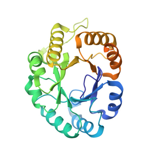
1U5H

Structure of Citrate Lyase beta subunit from Mycobacterium tuberculosis


Experimental Data Snapshot

  • Method: X-RAY DIFFRACTION
  • Resolution: 1.65 Å
  • R-Value Free: 0.272 
  • R-Value Work: 0.268 

wwPDB Validation   3D Report Full Report


This is version 1.4 of the entry. See complete history


Literature

The structure and computational analysis of Mycobacterium tuberculosis protein CitE suggest a novel enzymatic function.

Goulding, C.W.Bowers, P.M.Segelke, B.Lekin, T.Kim, C.Y.Terwilliger, T.C.Eisenberg, D.

(2007) J Mol Biol 365: 275-283

  • DOI: https://doi.org/10.1016/j.jmb.2006.09.086
  • Primary Citation of Related Structures:  
    1U5H, 1Z6K

  • PubMed Abstract: 

    Fatty acid biosynthesis is essential for the survival of Mycobacterium tuberculosis and acetyl-coenzyme A (acetyl-CoA) is an essential precursor in this pathway. We have determined the 3-D crystal structure of M. tuberculosis citrate lyase beta-subunit (CitE), which as annotated should cleave protein bound citryl-CoA to oxaloacetate and a protein-bound CoA derivative. The CitE structure has the (beta/alpha)(8) TIM barrel fold with an additional alpha-helix, and is trimeric. We have determined the ternary complex bound with oxaloacetate and magnesium, revealing some of the conserved residues involved in catalysis. While the bacterial citrate lyase is a complex with three subunits, the M. tuberculosis genome does not contain the alpha and gamma subunits of this complex, implying that M. tuberculosis CitE acts differently from other bacterial CitE proteins. The analysis of gene clusters containing the CitE protein from 168 fully sequenced organisms has led us to identify a grouping of functionally related genes preserved in M. tuberculosis, Rattus norvegicus, Homo sapiens, and Mus musculus. We propose a novel enzymatic function for M. tuberculosis CitE in fatty acid biosynthesis that is analogous to bacterial citrate lyase but producing acetyl-CoA rather than a protein-bound CoA derivative.


  • Organizational Affiliation

    Institute for Genomics and Proteomics, UCLA, Los Angeles, CA 90095-1570, USA.


Macromolecules
Find similar proteins by:  (by identity cutoff)  |  3D Structure
Entity ID: 1
MoleculeChains Sequence LengthOrganismDetailsImage
citE273Mycobacterium tuberculosisMutation(s): 5 
Gene Names: Rv2498c
EC: 4.1.3.6 (PDB Primary Data), 4.1 (UniProt)
UniProt
Find proteins for P9WPE1 (Mycobacterium tuberculosis (strain ATCC 25618 / H37Rv))
Explore P9WPE1 
Go to UniProtKB:  P9WPE1
Entity Groups  
Sequence Clusters30% Identity50% Identity70% Identity90% Identity95% Identity100% Identity
UniProt GroupP9WPE1
Sequence Annotations
Expand
  • Reference Sequence
Small Molecules
Ligands 1 Unique
IDChains Name / Formula / InChI Key2D Diagram3D Interactions
FMT
Query on FMT

Download Ideal Coordinates CCD File 
B [auth A]FORMIC ACID
C H2 O2
BDAGIHXWWSANSR-UHFFFAOYSA-N
Experimental Data & Validation

Experimental Data

  • Method: X-RAY DIFFRACTION
  • Resolution: 1.65 Å
  • R-Value Free: 0.272 
  • R-Value Work: 0.268 
  • Space Group: H 3 2
Unit Cell:
Length ( Å )Angle ( ˚ )
a = 91.214α = 90
b = 91.214β = 90
c = 221.233γ = 120
Software Package:
Software NamePurpose
CNSrefinement
DENZOdata reduction
SCALEPACKdata scaling
MLPHAREphasing

Structure Validation

View Full Validation Report


Entry History 

Deposition Data

Revision History  (Full details and data files)

  • Version 1.0: 2004-10-12
    Type: Initial release
  • Version 1.1: 2008-04-30
    Changes: Version format compliance
  • Version 1.2: 2011-07-13
    Changes: Version format compliance
  • Version 1.3: 2021-10-20
    Changes: Database references, Derived calculations
  • Version 1.4: 2024-02-14
    Changes: Data collection