1UMK

The Structure of Human Erythrocyte NADH-cytochrome b5 Reductase


Experimental Data Snapshot

  • Method: X-RAY DIFFRACTION
  • Resolution: 1.75 Å
  • R-Value Free: 
    0.207 (Depositor), 0.210 (DCC) 
  • R-Value Work: 
    0.165 (Depositor), 0.170 (DCC) 
  • R-Value Observed: 
    0.167 (Depositor) 

wwPDB Validation   3D Report Full Report


Ligand Structure Quality Assessment 

Created with Raphaël 2.3.0Worse 01 BetterLigand structure goodness of fit to experimental dataBest fitted FADClick on this verticalbar to view details

This is version 1.3 of the entry. See complete history


Literature

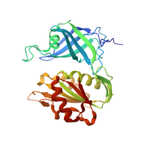
Structure of human erythrocyte NADH-cytochrome b5 reductase.

Bando, S.Takano, T.Yubisui, T.Shirabe, K.Takeshita, M.Nakagawa, A.

(2004) Acta Crystallogr D Biol Crystallogr 60: 1929-1934

  • DOI: https://doi.org/10.1107/S0907444904020645
  • Primary Citation of Related Structures:  
    1UMK

  • PubMed Abstract: 

    Erythrocyte NADH-cytochrome b(5) reductase reduces methaemoglobin to functional haemoglobin. In order to examine the function of the enzyme, the structure of NADH-cytochrome b(5) reductase from human erythrocytes has been determined and refined by X-ray crystallography. At 1.75 A resolution, the root-mean-square deviations (r.m.s.d.) from standard bond lengths and angles are 0.006 A and 1.03 degrees , respectively. The molecular structure was compared with those of rat NADH-cytochrome b(5) reductase and corn nitrate reductase. The human reductase resembles the rat reductase in overall structure as well as in many side chains. Nevertheless, there is a large main-chain shift from the human reductase to the rat reductase or the corn reductase caused by a single-residue replacement from proline to threonine. A model of the complex between cytochrome b(5) and the human reductase has been built and compared with that of the haem-containing domain of the nitrate reductase molecule. The interaction between cytochrome b(5) and the human reductase differs from that of the nitrate reductase because of differences in the amino-acid sequences. The structures around 15 mutation sites of the human reductase have been examined for the influence of residue substitutions using the program ROTAMER. Five mutations in the FAD-binding domain seem to be related to cytochrome b(5).


  • Organizational Affiliation

    Institute for Protein Research, Osaka University, 3-2 Yamadaoka, Suita, Osaka 565-0871, Japan.


Macromolecules
Find similar proteins by:  (by identity cutoff)  |  3D Structure
Entity ID: 1
MoleculeChains Sequence LengthOrganismDetailsImage
NADH-cytochrome b5 reductase275Homo sapiensMutation(s): 0 
EC: 1.6.2.2
UniProt & NIH Common Fund Data Resources
Find proteins for P00387 (Homo sapiens)
Explore P00387 
Go to UniProtKB:  P00387
PHAROS:  P00387
GTEx:  ENSG00000100243 
Entity Groups  
Sequence Clusters30% Identity50% Identity70% Identity90% Identity95% Identity100% Identity
UniProt GroupP00387
Sequence Annotations
Expand
  • Reference Sequence
Small Molecules
Ligands 1 Unique
IDChains Name / Formula / InChI Key2D Diagram3D Interactions
FAD
Query on FAD

Download Ideal Coordinates CCD File 
B [auth A]FLAVIN-ADENINE DINUCLEOTIDE
C27 H33 N9 O15 P2
VWWQXMAJTJZDQX-UYBVJOGSSA-N
Experimental Data & Validation

Experimental Data

  • Method: X-RAY DIFFRACTION
  • Resolution: 1.75 Å
  • R-Value Free:  0.207 (Depositor), 0.210 (DCC) 
  • R-Value Work:  0.165 (Depositor), 0.170 (DCC) 
  • R-Value Observed: 0.167 (Depositor) 
Space Group: P 41
Unit Cell:
Length ( Å )Angle ( ˚ )
a = 65.869α = 90
b = 65.869β = 90
c = 76.2γ = 90
Software Package:
Software NamePurpose
REFMACrefinement
MOSFLMdata reduction
CCP4data scaling
MLPHAREphasing

Structure Validation

View Full Validation Report


Ligand Structure Quality Assessment 

Created with Raphaël 2.3.0Worse 01 BetterLigand structure goodness of fit to experimental dataBest fitted FADClick on this verticalbar to view details

Entry History 

Deposition Data

Revision History  (Full details and data files)

  • Version 1.0: 2004-11-02
    Type: Initial release
  • Version 1.1: 2008-04-27
    Changes: Version format compliance
  • Version 1.2: 2011-07-13
    Changes: Version format compliance
  • Version 1.3: 2023-12-27
    Changes: Data collection, Database references, Derived calculations