(1R,4S,5R)-3-Fluoro-1,4,5-Trihydroxy-2-Cyclohexene-1-Carboxylic Acid: The Fluoro Analogue of the Enolate Intermediate in the Reaction Catalyzed by Type II Dehydroquinases
Frederickson, M., Roszak, A.W., Coggins, J.R., Lapthorn, A.J., Abell, C.(2004) Org Biomol Chem 2: 1592
- PubMed: 15162210 Search on PubMed
- DOI: https://doi.org/10.1039/b404535a
- Primary Citation Related Structures:
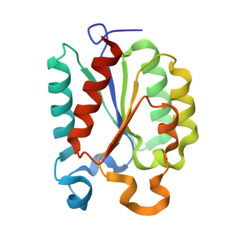
1V1J - PubMed Abstract:
The fluoro analogue of the enolate intermediate in the reaction catalyzed by type II dehydroquinases has been prepared from naturally occurring (-)-quinic acid over seven steps and has been shown to be the most potent inhibitor reported to date of the type II enzyme from Mycobacterium tuberculosis.
- University Chemical Laboratory, Lensfield Road, Cambridge, UK CB2 1EW.
Organizational Affiliation: