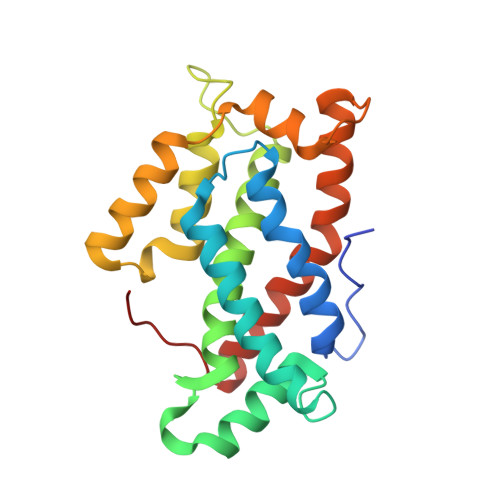
1WBE

X-ray structure of bovine GLTP


Experimental Data Snapshot

  • Method: X-RAY DIFFRACTION
  • Resolution: 1.36 Å
  • R-Value Free: 0.219 
  • R-Value Work: 0.184 
  • R-Value Observed: 0.186 

Starting Model: experimental
View more details

wwPDB Validation   3D Report Full Report


Ligand Structure Quality Assessment 


This is version 1.4 of the entry. See complete history


Literature

Structural Evidence for Adaptive Ligand Binding of Glycolipid Transfer Protein.

Airenne, T.T.Kidron, H.Nymalm, Y.Nylund, M.West, G.Mattjus, P.Salminen, T.A.

(2006) J Mol Biol 355: 224

  • DOI: https://doi.org/10.1016/j.jmb.2005.10.031
  • Primary Citation of Related Structures:  
    1TFJ, 1WBE, 2BV7

  • PubMed Abstract: 

    Glycolipids participate in many important cellular processes and they are bound and transferred with high specificity by glycolipid transfer protein (GLTP). We have solved three different X-ray structures of bovine GLTP at 1.4 angstroms, 1.6 angstroms and 1.8 angstroms resolution, all with a bound fatty acid or glycolipid. The 1.4 angstroms structure resembles the recently characterized apo-form of the human GLTP but the other two structures represent an intermediate conformation of the apo-GLTPs and the human lactosylceramide-bound GLTP structure. These novel structures give insight into the mechanism of lipid binding and how GLTP may conformationally adapt to different lipids. Furthermore, based on the structural comparison of the GLTP structures and the three-dimensional models of the related Podospora anserina HET-C2 and Arabidopsis thaliana accelerated cell death protein, ACD11, we give structural explanations for their specific lipid binding properties.


  • Organizational Affiliation

    Department of Biochemistry and Pharmacy, Abo Akademi University, Tykistökatu 6A, FIN-20520 Turku, Finland.


Macromolecules
Find similar proteins by:  (by identity cutoff)  |  3D Structure
Entity ID: 1
MoleculeChains Sequence LengthOrganismDetailsImage
GLYCOLIPID TRANSFER PROTEIN209Bos taurusMutation(s): 0 
UniProt
Find proteins for P68265 (Bos taurus)
Explore P68265 
Go to UniProtKB:  P68265
Entity Groups  
Sequence Clusters30% Identity50% Identity70% Identity90% Identity95% Identity100% Identity
UniProt GroupP68265
Sequence Annotations
Expand
  • Reference Sequence
Small Molecules
Ligands 2 Unique
IDChains Name / Formula / InChI Key2D Diagram3D Interactions
DKA
Query on DKA

Download Ideal Coordinates CCD File 
B [auth A]DECANOIC ACID
C10 H20 O2
GHVNFZFCNZKVNT-UHFFFAOYSA-N
GOL
Query on GOL

Download Ideal Coordinates CCD File 
C [auth A]GLYCEROL
C3 H8 O3
PEDCQBHIVMGVHV-UHFFFAOYSA-N
Experimental Data & Validation

Experimental Data

  • Method: X-RAY DIFFRACTION
  • Resolution: 1.36 Å
  • R-Value Free: 0.219 
  • R-Value Work: 0.184 
  • R-Value Observed: 0.186 
  • Space Group: P 1 21 1
Unit Cell:
Length ( Å )Angle ( ˚ )
a = 55.741α = 90
b = 35.393β = 116.55
c = 58.423γ = 90
Software Package:
Software NamePurpose
REFMACrefinement
XDSdata reduction
XDSdata scaling
ARP/wARPphasing

Structure Validation

View Full Validation Report


Ligand Structure Quality Assessment 


Entry History 

Deposition Data

Revision History  (Full details and data files)

  • Version 1.0: 2005-11-30
    Type: Initial release
  • Version 1.1: 2011-05-08
    Changes: Version format compliance
  • Version 1.2: 2011-07-13
    Changes: Version format compliance
  • Version 1.3: 2017-06-28
    Changes: Refinement description
  • Version 1.4: 2023-12-13
    Changes: Data collection, Database references, Other, Refinement description