Structural basis for mRNA Cap-Binding regulation of eukaryotic initiation factor 4E by 4E-binding protein, studied by spectroscopic, X-ray crystal structural, and molecular dynamics simulation methods
Tomoo, K., Matsushita, Y., Fujisaki, H., Abiko, F., Shen, X., Taniguchi, T., Miyagawa, H., Kitamura, K., Miura, K., Ishida, T.(2005) Biochim Biophys Acta 1753: 191-208
- PubMed: 16271312 Search on PubMed
- DOI: https://doi.org/10.1016/j.bbapap.2005.07.023
- Primary Citation Related Structures:
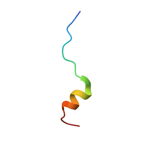
1WKW - PubMed Abstract:
Taking advantage of the Trp73 residue located close to the 4E-BP binding site of eIF4E, the interaction between the 4E-BP isoform and eIF4E was investigated by the Trp fluorescence titration method. Although no significant difference was observed among the association constants of three 4E-BP isoforms, the binding preference of 4E-BP2 over 4E-BP1 and -BP3 was shown, probably due to the effect of a 4E-BP2-specific LDRR (60-63) sequence for the binding with eIF4E. By contrast, surface plasmon resonance (SPR) analyses showed the binding preference of 4E-BP1, although the difference among the isoforms was also not significant. This inconsistency with fluorescence analysis likely resulted from the different observation points of the interaction, i.e., local and overall interactions observed by the fluorescence and SPR methods, respectively. To clarify the structural basis for these spectroscopic results, the crystal structure of the ternary complex of m7GpppA-eIF4E-4E-BP1 fragment (Thr36-Thr70) was analyzed by the X-ray diffraction method. Crystal structure analysis at 2.1 A resolution revealed that the 4E-BP1 fragment, assigned to the Pro47-Pro66 peptide moiety, adopted a reverse L-shaped conformation involving the beta sheet and alpha-helical structures and was located at the root of the handle of the temple-bell-shaped eIF4E through hydrophilic and hydrophobic interactions. Based on the observed binding mode, possible interactions with the three 4E-BP isoforms have been discussed. On the other hand, since the crystal structural comparison with the previously determined m7GpppA-eIF4E-4E binary complex showed that the docking of the 4E-BP1 fragment does not significantly affect the overall tertiary structure and cap-binding scaffold of eIF4E, the dynamic regulation of the cap-binding of eIF4E by 4E-BP1 was investigated by molecular dynamics (MD) simulations. Consequently, the simulation suggested that (i) the helical region of the 4E-BP1 peptide is important for the binding with eIF4E, (ii) the existence of a cap structure stabilizes the binding of eIF4E with 4E-BP, (iii) the binding of 4E-BP stabilizes the cap-binding pocket of eIF4E, and (iv) the phosphorylation of Ser67 alone does not induce the separation of 4E-BP from eIF4E, but increases the structural rigidity of 4E-BP. These results provide the structural basis for the mRNA cap-binding regulation of eIF4E by 4E-BP.
- Department of Physical Chemistry, Osaka University of Pharmaceutical Sciences, 4-20-1 Nasahara, Takatsuki, Osaka 569-1094, Japan. tomoo@gly.oups.ac.jp
Organizational Affiliation: