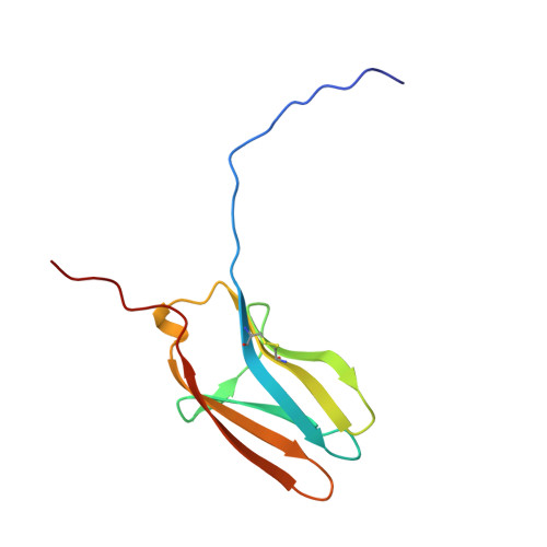
Crystal structures of the neurotrophin-binding domain of TrkA, TrkB and TrkC.
Ultsch, M.H., Wiesmann, C., Simmons, L.C., Henrich, J., Yang, M., Reilly, D., Bass, S.H., de Vos, A.M.(1999) J Mol Biol 290: 149-159
- PubMed: 10388563 Search on PubMed
- DOI: https://doi.org/10.1006/jmbi.1999.2816
- Primary Citation Related Structures:
1WWA, 1WWB, 1WWC - PubMed Abstract:
The Trk receptors and their neurotrophin ligands control development and maintenance of the nervous system. The crystal structures of the ligand binding domain of TrkA, TrkB, and TrkC were solved and refined to high resolution. The domains adopt an immunoglobulin-like fold, but crystallized in all three instances as dimers with the N-terminal strand of each molecule replaced by the same strand of a symmetry-related mate. Models of the correctly folded domains could be constructed by changing the position of a single residue, and the resulting model of the binding domain of TrkA is essentially identical with the bound structure as observed in a complex with nerve growth factor. An analysis of the existing mutagenesis data for TrkA and TrkC in light of these structures reveals the structural reasons for the specificity among the Trk receptors, and explains the underpinnings of the multi-functional ligands that have been reported. The overall structure of all three domains belongs to the I-set of immunoglobulin-like domains, but shows several unusual features, such as an exposed disulfide bridge linking two neighboring strands in the same beta-sheet. For all three domains, the residues that deviate from the standard fingerprint pattern common to the I-set family fall in the region of the ligand binding site observed in the complex. Therefore, identification of these deviations in the sequences of other immunoglobulin-like domain-containing receptors may help to identify their ligand binding site even in the absence of structural or mutagenesis data.
- Department of Protein Engineering, Genentech, Inc., South San Francisco, CA 94080, USA.
Organizational Affiliation: