1X0S | pdb_00001x0s

Crystal structure of the 13-cis isomer of bacteriorhodopsin


Experimental Data Snapshot

  • Method: X-RAY DIFFRACTION
  • Resolution: 2.50 Å
  • R-Value Free: 
    0.305 (Depositor), 0.317 (DCC) 
  • R-Value Work: 
    0.271 (Depositor), 0.283 (DCC) 
  • R-Value Observed: 
    0.271 (Depositor) 

Starting Model: experimental
View more details

wwPDB Validation 3D Report Full Report

Validation slider image for 1X0S

Ligand Structure Quality Assessment 


This is version 2.2 of the entry. See complete history

Literature

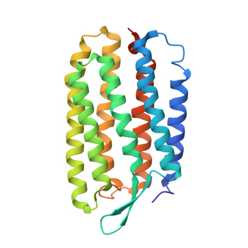
Crystal structure of the 13-cis isomer of bacteriorhodopsin in the dark-adapted state.

Nishikawa, T.Murakami, M.Kouyama, T.

(2005) J Mol Biology 352: 319-328

  • DOI: https://doi.org/10.1016/j.jmb.2005.07.021
  • Primary Citation Related Structures: 
    1X0S

  • PubMed Abstract: 

    The atomic structure of the trans isomer of bacteriorhodopsin was determined previously by using a 3D crystal belonging to the space group P622. Here, a structure is reported for another isomer with the 13-cis, 15-syn retinal in a dark-adapted crystal. Structural comparison of the two isomers indicates that retinal isomerization around the C13[double bond]C14 and the C15[double bond]N bonds is accompanied by noticeable displacements of a few residues in the vicinity of the retinal Schiff base and small re-arrangement of the hydrogen-bonding network in the proton release channel. On the other hand, aromatic residues surrounding the retinal polyene chain were found to scarcely move during the dark/light adaptation. This result suggests that variation in the structural rigidity within the retinal-binding pocket is one of the important factors ensuring the stereospecific isomerization of retinal.


  • Organizational Affiliation
    • Department of Physics, Graduate School of Science, Nagoya University, Nagoya 464-8602, Japan.

Macromolecule Content 

  • Total Structure Weight: 31.89 kDa 
  • Atom Count: 2,101 
  • Modeled Residue Count: 227 
  • Deposited Residue Count: 248 
  • Unique protein chains: 1

Macromolecules

Find similar proteins by:|  3D Structure
Entity ID: 1
MoleculeChains  Sequence LengthOrganismDetailsImage
Bacteriorhodopsin248Halobacterium salinarumMutation(s): 0 
Membrane Entity: Yes 
UniProt
Find proteins for P02945 (Halobacterium salinarum (strain ATCC 700922 / JCM 11081 / NRC-1))
Explore P02945 
Go to UniProtKB:  P02945
Entity Groups
Sequence Clusters30% Identity50% Identity70% Identity90% Identity95% Identity100% Identity
UniProt GroupP02945
Sequence Annotations
Expand
Reference Sequence

Oligosaccharides

Help  
Entity ID: 2
MoleculeChains Length2D Diagram GlycosylationD Interactions
beta-D-galactopyranose-(1-6)-alpha-D-mannopyranose-(1-2)-alpha-D-glucopyranose
B
3N/A
Glycosylation Resources
GlyTouCan: G59827MO
GlyCosmos: G59827MO

Small Molecules

Ligands 4 Unique
IDChains Name / Formula / InChI Key2D Diagram3D Interactions
L3P

Query on L3P


Download:Ideal Coordinates CCD File
E [auth A],
G [auth A],
H [auth A],
I [auth A]
2,3-DI-O-PHYTANLY-3-SN-GLYCERO-1-PHOSPHORYL-3'-SN-GLYCEROL-1'-PHOSPHATE
C46 H94 O11 P2
TZXJQSKPTCRGCA-VZSPAKCESA-L
L2P

Query on L2P


Download:Ideal Coordinates CCD File
F [auth A]2,3-DI-PHYTANYL-GLYCEROL
C43 H88 O3
ISDBCJSGCHUHFI-UMZPFTBHSA-N
RET

Query on RET


Download:Ideal Coordinates CCD File
D [auth A]RETINAL
C20 H28 O
NCYCYZXNIZJOKI-OVSJKPMPSA-N
SO4

Query on SO4


Download:Ideal Coordinates CCD File
C [auth A]SULFATE ION
O4 S
QAOWNCQODCNURD-UHFFFAOYSA-L

Experimental Data & Validation

Experimental Data

  • Method: X-RAY DIFFRACTION
  • Resolution: 2.50 Å
  • R-Value Free:  0.305 (Depositor), 0.317 (DCC) 
  • R-Value Work:  0.271 (Depositor), 0.283 (DCC) 
  • R-Value Observed: 0.271 (Depositor) 
Space Group: P 6 2 2
Unit Cell:
Length ( Å )Angle ( ˚ )
a = 101.98α = 90
b = 101.98β = 90
c = 112.1γ = 120
Software Package:
Software NamePurpose
CNSrefinement
MOSFLMdata reduction
CCP4data scaling
XTALVIEWrefinement

Structure Validation

View Full Validation Report


Ligand Structure Quality Assessment 


Entry History 

Deposition Data

Revision History  (Full details and data files)

  • Version 1.0: 2005-08-30
    Type: Initial release
  • Version 1.1: 2008-04-30
    Changes: Version format compliance
  • Version 1.2: 2011-07-13
    Changes: Derived calculations, Version format compliance
  • Version 1.3: 2019-11-06
    Changes: Data collection, Derived calculations
  • Version 2.0: 2020-07-29
    Type: Remediation
    Reason: Carbohydrate remediation
    Changes: Advisory, Atomic model, Data collection, Derived calculations, Structure summary
  • Version 2.1: 2023-10-25
    Changes: Data collection, Database references, Derived calculations, Refinement description, Structure summary
  • Version 2.2: 2024-10-23
    Changes: Structure summary