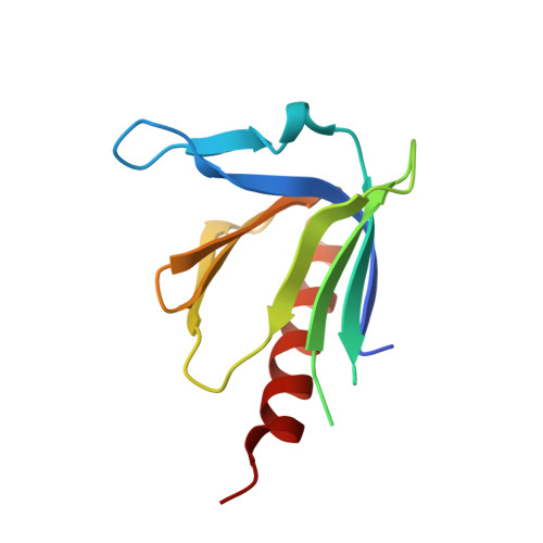
1.15A Crystal structure of the X. tropicalis Spred1 EVH1 domain suggests a fourth distinct peptide-binding mechanism within the EVH1 family
Harmer, N.J., Sivak, J.M., Amaya, E., Blundell, T.L.(2005) FEBS Lett 579: 1161-1166
- PubMed: 15710406 Search on PubMed
- DOI: https://doi.org/10.1016/j.febslet.2004.11.114
- Primary Citation Related Structures:
1XOD - PubMed Abstract:
The recently described Spred protein family has been implicated in the modulation of receptor tyrosine kinase signalling. We report the crystal structure of the Enabled/vasodilator-stimulated phosphoprotein homology-1 (EVH1) domain from Xenopus tropicalis Spred1, solved to 1.15 A resolution. This structure confirms that the Spred EVH1 adopts the pleckstrin-homology fold, with a similar secondary structure to Enabled. A translation of one of the peptide-binding groove beta-strands narrows this groove, whilst one end of the groove shows structural flexibility. We propose that Spred1 will bind peptides that are less proline-rich than other EVH1 domains, with conformational changes indicating an induced fit.
- Department of Biochemistry, 80 Tennis Court Road, Cambridge, CB2 1GA, UK. nic@cryst.bioc.cam.ac.uk
Organizational Affiliation: