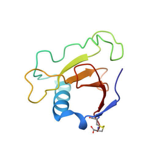
Asp79 Makes a Large, Unfavorable Contribution to the Stability of RNase Sa.
Trevino, S.R., Gokulan, K., Newsom, S., Thurlkill, R.L., Shaw, K.L., Mitkevich, V.A., Makarov, A.A., Sacchettini, J.C., Scholtz, J.M., Pace, C.N.(2005) J Mol Biol 354: 967-978
- PubMed: 16288913
- DOI: https://doi.org/10.1016/j.jmb.2005.09.091
- Primary Citation of Related Structures:
1YNV - PubMed Abstract:
The two most buried carboxyl groups in ribonuclease Sa (RNase Sa) are Asp33 (99% buried; pK 2.4) and Asp79 (85% buried; pK 7.4). Above these pK values, the stability of the D33A variant is 6kcal/mol less than wild-type RNase Sa, and the stability of the D79A variant is 3.3kcal/mol greater than wild-type RNase Sa. The key structural difference between the carboxyl groups is that Asp33 forms three intramolecular hydrogen bonds, and Asp79 forms no intramolecular hydrogen bond. Here, we focus on Asp79 and describe studies of 11 Asp79 variants. Most of the variants were at least 2kcal/mol more stable than wild-type RNase Sa, and the most interesting was D79F. At pH 3, below the pK of Asp79, RNase Sa is 0.3kcal/mol more stable than the D79F variant. At pH 8.5, above the pK of Asp79, RNase Sa is 3.7kcal/mol less stable than the D79F variant. The unfavorable contribution of Asp79 to the stability appears to result from the Born self-energy of burying the charge and, more importantly, from unfavorable charge-charge interactions. To counteract the effect of the negative charge on Asp79, we prepared the Q94K variant and the crystal structure showed that the amino group of the Lys formed a hydrogen-bonded ion pair (distance, 2.71A; angle, 100 degrees ) with the carboxyl group of Asp79. The stability of the Q94K variant was about the same as the wild-type at pH 3, where Asp79 is uncharged, but 1kcal/mol greater than that of wild-type RNase Sa at pH 8.5, where Asp79 is charged. Differences in hydrophobicity, steric strain, Born self-energy, and electrostatic interactions all appear to contribute to the range of stabilities observed in the variants. When it is possible, replacing buried, non-hydrogen bonded, ionizable side-chains with non-polar side-chains is an excellent means of increasing protein stability.
Organizational Affiliation:
Department of Medical Biochemistry and Genetics, Texas A and M University, College Station, TX 77843, USA.


