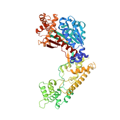
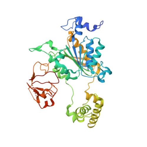
1YOV

Insights into the Ubiquitin Transfer Cascade from the refined structure of the activating enzyme for NEDD8


Experimental Data Snapshot

  • Method: X-RAY DIFFRACTION
  • Resolution: 2.60 Å
  • R-Value Free: 0.280 
  • R-Value Work: 0.236 

wwPDB Validation   3D Report Full Report


This is version 1.3 of the entry. See complete history


Literature

Insights into the Ubiquitin Transfer Cascade from the refined structure of the activating enzyme for NEDD8

Walden, H.Podgorski, M.S.Schulman, B.A.

(2003) Nature 422: 330-334

  • DOI: https://doi.org/10.1038/nature01456
  • Primary Citation of Related Structures:  
    1YOV

  • PubMed Abstract: 

    Post-translational modification by ubiquitin-like proteins (Ublps) is an essential cellular regulatory mechanism. The Ublp NEDD8 regulates cell division, signalling and embryogenesis. Ublps are conjugated to their targets by the sequential action of E1, E2 and often E3 enzymes. Each Ublp has a dedicated E1, or activating enzyme, that initiates its conjugation cascade. First, E1 associates with the Ublp and catalyses adenylation of the carboxy terminus of the Ublp. Second, E1 forms a thioester between its catalytic cysteine and the Ublp. Next, E1 is loaded with a second Ublp molecule, adenylating the C terminus of this second Ublp while still carrying the first thioester-bound Ublp. Last, E1 binds E2 and promotes Ublp transfer to the catalytic cysteine of E2. We report here the structure and mutational analysis of human APPBP1-UBA3, the heterodimeric E1 enzyme for NEDD8 (ref. 11). Each E1 activity is specified by a domain: an adenylation domain resembling bacterial adenylating enzymes, an E1-specific domain organized around the catalytic cysteine, and a domain involved in E2 recognition resembling ubiquitin. The domains are arranged around two clefts that coordinate protein and nucleotide binding so that each of E1's reactions drives the next, in an assembly-line fashion.


  • Organizational Affiliation

    Department of Structural Biology, St Jude Children's Research Hospital, Memphis, Tennessee 38105, USA.


Macromolecules
Find similar proteins by:  (by identity cutoff)  |  3D Structure
Entity ID: 1
MoleculeChains Sequence LengthOrganismDetailsImage
Amyloid protein-binding protein 1
A, C
537Homo sapiensMutation(s): 0 
Gene Names: APPBP1
UniProt & NIH Common Fund Data Resources
Find proteins for Q13564 (Homo sapiens)
Explore Q13564 
Go to UniProtKB:  Q13564
PHAROS:  Q13564
GTEx:  ENSG00000159593 
Entity Groups  
Sequence Clusters30% Identity50% Identity70% Identity90% Identity95% Identity100% Identity
UniProt GroupQ13564
Sequence Annotations
Expand
  • Reference Sequence
Find similar proteins by:  (by identity cutoff)  |  3D Structure
Entity ID: 2
MoleculeChains Sequence LengthOrganismDetailsImage
Ubiquitin-activating enzyme E1C
B, D
444Homo sapiensMutation(s): 0 
Gene Names: UBE1CUBA3
EC: 6.2.1.64
UniProt & NIH Common Fund Data Resources
Find proteins for Q8TBC4 (Homo sapiens)
Explore Q8TBC4 
Go to UniProtKB:  Q8TBC4
PHAROS:  Q8TBC4
GTEx:  ENSG00000144744 
Entity Groups  
Sequence Clusters30% Identity50% Identity70% Identity90% Identity95% Identity100% Identity
UniProt GroupQ8TBC4
Sequence Annotations
Expand
  • Reference Sequence
Experimental Data & Validation

Experimental Data

  • Method: X-RAY DIFFRACTION
  • Resolution: 2.60 Å
  • R-Value Free: 0.280 
  • R-Value Work: 0.236 
  • Space Group: P 21 21 21
Unit Cell:
Length ( Å )Angle ( ˚ )
a = 92.4α = 90
b = 123.6β = 90
c = 198.2γ = 90
Software Package:
Software NamePurpose
DENZOdata reduction
SCALEPACKdata scaling
SOLVEphasing
CNSrefinement

Structure Validation

View Full Validation Report


Entry History 

Deposition Data

Revision History  (Full details and data files)

  • Version 1.0: 2005-03-08
    Type: Initial release
  • Version 1.1: 2008-04-30
    Changes: Version format compliance
  • Version 1.2: 2011-07-13
    Changes: Version format compliance
  • Version 1.3: 2024-02-14
    Changes: Data collection, Database references, Derived calculations