1YSL | pdb_00001ysl

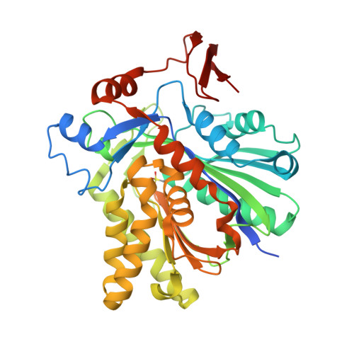
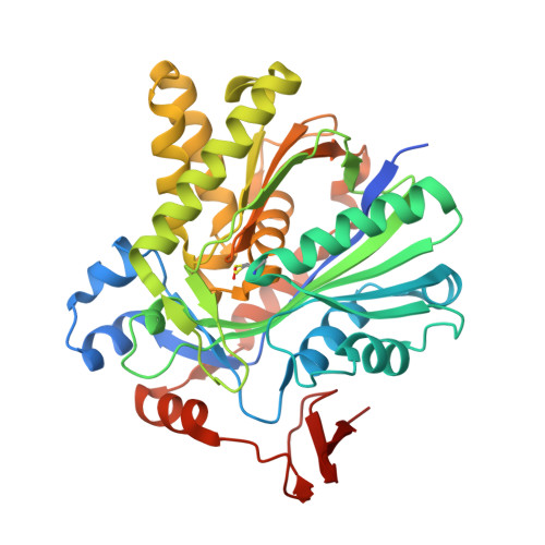
Crystal structure of HMG-CoA synthase from Enterococcus faecalis with AcetoAcetyl-CoA ligand.


Experimental Data Snapshot

  • Method: X-RAY DIFFRACTION
  • Resolution: 1.90 Å
  • R-Value Free: 
    0.201 (Depositor), 0.199 (DCC) 
  • R-Value Work: 
    0.179 (Depositor), 0.176 (DCC) 
  • R-Value Observed: 
    0.180 (Depositor) 

Starting Model: experimental
View more details

wwPDB Validation   3D Report Full Report


Ligand Structure Quality Assessment 

Created with Raphaël 2.3.0Worse 01 BetterLigand structure goodness of fit to experimental dataBest fitted COAClick on this verticalbar to view details

This is version 1.5 of the entry. See complete history


Literature

X-ray Crystal Structures of HMG-CoA Synthase from Enterococcus faecalis and a Complex with Its Second Substrate/Inhibitor Acetoacetyl-CoA.

Steussy, C.N.Vartia, A.A.Burgner II, J.W.Sutherlin, A.Rodwell, V.W.Stauffacher, C.V.

(2005) Biochemistry 44: 14256-14267

  • DOI: https://doi.org/10.1021/bi051487x
  • Primary Citation of Related Structures:  
    1X9E, 1YSL

  • PubMed Abstract: 

    Biosynthesis of the isoprenoid precursor, isopentenyl diphosphate, is a critical function in all independently living organisms. There are two major pathways for this synthesis, the non-mevalonate pathway found in most eubacteria and the mevalonate pathway found in animal cells and a number of pathogenic bacteria. An early step in this pathway is the condensation of acetyl-CoA and acetoacetyl-CoA into HMG-CoA, catalyzed by the enzyme HMG-CoA synthase. To explore the possibility of a small molecule inhibitor of the enzyme functioning as a non-cell wall antibiotic, the structure of HMG-CoA synthase from Enterococcus faecalis (MVAS) was determined by selenomethionine MAD phasing to 2.4 A and the enzyme complexed with its second substrate, acetoacetyl-CoA, to 1.9 A. These structures show that HMG-CoA synthase from Enterococcus is a member of the family of thiolase fold enzymes and, while similar to the recently published HMG-CoA synthase structures from Staphylococcus aureus, exhibit significant differences in the structure of the C-terminal domain. The acetoacetyl-CoA binary structure demonstrates reduced coenzyme A and acetoacetate covalently bound to the active site cysteine through a thioester bond. This is consistent with the kinetics of the reaction that have shown acetoacetyl-CoA to be a potent inhibitor of the overall reaction, and provides a starting point in the search for a small molecule inhibitor.


  • Organizational Affiliation

    Department of Biological Sciences, Purdue University, West Lafayette, Indiana 47907, USA.


Macromolecules
Find similar proteins by:  (by identity cutoff)  |  3D Structure
Entity ID: 1
MoleculeChains Sequence LengthOrganismDetailsImage
HMG-CoA synthase402Enterococcus faecalisMutation(s): 0 
EC: 4.1.3.5 (PDB Primary Data), 2.3.3.10 (UniProt)
UniProt
Find proteins for Q9FD71 (Enterococcus faecalis)
Explore Q9FD71 
Go to UniProtKB:  Q9FD71
Entity Groups  
Sequence Clusters30% Identity50% Identity70% Identity90% Identity95% Identity100% Identity
UniProt GroupQ9FD71
Sequence Annotations
Expand
  • Reference Sequence
Find similar proteins by:  (by identity cutoff)  |  3D Structure
Entity ID: 2
MoleculeChains Sequence LengthOrganismDetailsImage
HMG-CoA synthase402Enterococcus faecalisMutation(s): 1 
EC: 4.1.3.5 (PDB Primary Data), 2.3.3.10 (UniProt)
UniProt
Find proteins for Q9FD71 (Enterococcus faecalis)
Explore Q9FD71 
Go to UniProtKB:  Q9FD71
Entity Groups  
Sequence Clusters30% Identity50% Identity70% Identity90% Identity95% Identity100% Identity
UniProt GroupQ9FD71
Sequence Annotations
Expand
  • Reference Sequence
Small Molecules
Ligands 4 Unique
IDChains Name / Formula / InChI Key2D Diagram3D Interactions
COA
Query on COA

Download Ideal Coordinates CCD File 
F [auth B]COENZYME A
C21 H36 N7 O16 P3 S
RGJOEKWQDUBAIZ-IBOSZNHHSA-N
AAE
Query on AAE

Download Ideal Coordinates CCD File 
G [auth B]ACETOACETIC ACID
C4 H6 O3
WDJHALXBUFZDSR-UHFFFAOYSA-N
SO4
Query on SO4

Download Ideal Coordinates CCD File 
C [auth A],
E [auth B]
SULFATE ION
O4 S
QAOWNCQODCNURD-UHFFFAOYSA-L
GOL
Query on GOL

Download Ideal Coordinates CCD File 
D [auth A],
H [auth B]
GLYCEROL
C3 H8 O3
PEDCQBHIVMGVHV-UHFFFAOYSA-N
Modified Residues  1 Unique
IDChains TypeFormula2D DiagramParent
CSD
Query on CSD
A
L-PEPTIDE LINKINGC3 H7 N O4 SCYS
Experimental Data & Validation

Experimental Data

  • Method: X-RAY DIFFRACTION
  • Resolution: 1.90 Å
  • R-Value Free:  0.201 (Depositor), 0.199 (DCC) 
  • R-Value Work:  0.179 (Depositor), 0.176 (DCC) 
  • R-Value Observed: 0.180 (Depositor) 
Space Group: I 2 2 2
Unit Cell:
Length ( Å )Angle ( ˚ )
a = 105.3α = 90
b = 109.013β = 90
c = 140.799γ = 90
Software Package:
Software NamePurpose
HKL-2000data collection
SCALEPACKdata scaling
CNSrefinement
HKL-2000data reduction
CNSphasing

Structure Validation

View Full Validation Report


Ligand Structure Quality Assessment 

Created with Raphaël 2.3.0Worse 01 BetterLigand structure goodness of fit to experimental dataBest fitted COAClick on this verticalbar to view details

Entry History 

Deposition Data

Revision History  (Full details and data files)

  • Version 1.0: 2005-11-08
    Type: Initial release
  • Version 1.1: 2008-04-30
    Changes: Version format compliance
  • Version 1.2: 2011-07-13
    Changes: Non-polymer description, Version format compliance
  • Version 1.3: 2011-11-16
    Changes: Atomic model
  • Version 1.4: 2023-08-23
    Changes: Data collection, Database references, Derived calculations, Refinement description, Structure summary
  • Version 1.5: 2024-12-25
    Changes: Advisory, Derived calculations, Structure summary