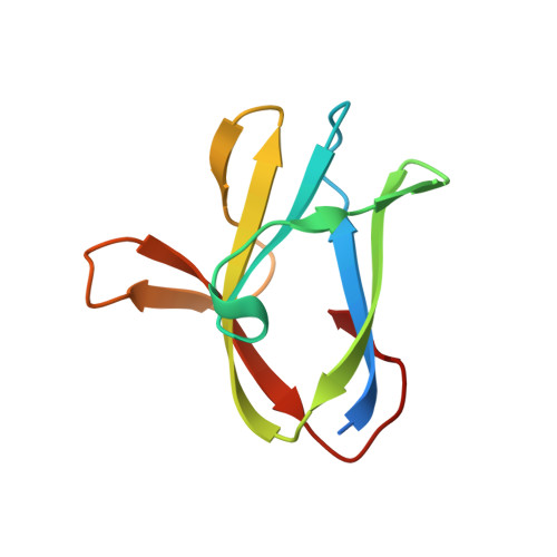
1Z0M

the glycogen-binding domain of the AMP-activated protein kinase beta1 subunit


Experimental Data Snapshot

  • Method: X-RAY DIFFRACTION
  • Resolution: 1.91 Å
  • R-Value Free: 0.243 
  • R-Value Work: 0.191 
  • R-Value Observed: 0.227 

wwPDB Validation   3D Report Full Report


This is version 2.1 of the entry. See complete history


Literature

Structural Basis for Glycogen Recognition by AMP-Activated Protein Kinase.

Polekhina, G.Gupta, A.van Denderen, B.J.Feil, S.C.Kemp, B.E.Stapleton, D.Parker, M.W.

(2005) Structure 13: 1453-1462

  • DOI: https://doi.org/10.1016/j.str.2005.07.008
  • Primary Citation of Related Structures:  
    1Z0M, 1Z0N

  • PubMed Abstract: 

    AMP-activated protein kinase (AMPK) coordinates cellular metabolism in response to energy demand as well as to a variety of stimuli. The AMPK beta subunit acts as a scaffold for the alpha catalytic and gamma regulatory subunits and targets the AMPK heterotrimer to glycogen. We have determined the structure of the AMPK beta glycogen binding domain in complex with beta-cyclodextrin. The structure reveals a carbohydrate binding pocket that consolidates all known aspects of carbohydrate binding observed in starch binding domains into one site, with extensive contact between several residues and five glucose units. beta-cyclodextrin is held in a pincer-like grasp with two tryptophan residues cradling two beta-cyclodextrin glucose units and a leucine residue piercing the beta-cyclodextrin ring. Mutation of key beta-cyclodextrin binding residues either partially or completely prevents the glycogen binding domain from binding glycogen. Modeling suggests that this binding pocket enables AMPK to interact with glycogen anywhere across the carbohydrate's helical surface.


  • Organizational Affiliation

    St. Vincent's Institute of Medical Research, 41 Victoria Parade, Fitzroy, Victoria 3065, Australia.


Macromolecules
Find similar proteins by:  (by identity cutoff)  |  3D Structure
Entity ID: 1
MoleculeChains Sequence LengthOrganismDetailsImage
5'-AMP-activated protein kinase, beta-1 subunit
A, B, C
96Rattus norvegicusMutation(s): 0 
UniProt
Find proteins for P80386 (Rattus norvegicus)
Explore P80386 
Go to UniProtKB:  P80386
Entity Groups  
Sequence Clusters30% Identity50% Identity70% Identity90% Identity95% Identity100% Identity
UniProt GroupP80386
Sequence Annotations
Expand
  • Reference Sequence
Oligosaccharides

Help

Entity ID: 2
MoleculeChains Length2D Diagram Glycosylation3D Interactions
Cycloheptakis-(1-4)-(alpha-D-glucopyranose)
D, E, F
7N/A
Glycosylation Resources
GlyTouCan:  G01435GL
GlyCosmos:  G01435GL
Biologically Interesting Molecules (External Reference) 1 Unique
Experimental Data & Validation

Experimental Data

  • Method: X-RAY DIFFRACTION
  • Resolution: 1.91 Å
  • R-Value Free: 0.243 
  • R-Value Work: 0.191 
  • R-Value Observed: 0.227 
  • Space Group: P 1
Unit Cell:
Length ( Å )Angle ( ˚ )
a = 43.735α = 70.72
b = 44.891β = 68.77
c = 50.335γ = 65.79
Software Package:
Software NamePurpose
DENZOdata reduction
SCALEPACKdata scaling
MLPHAREphasing
CNSrefinement
REFMACrefinement
PDB_EXTRACTdata extraction
DMphasing

Structure Validation

View Full Validation Report


Entry History 

Deposition Data

Revision History  (Full details and data files)

  • Version 1.0: 2005-10-25
    Type: Initial release
  • Version 1.1: 2008-04-30
    Changes: Version format compliance
  • Version 1.2: 2011-07-13
    Changes: Version format compliance
  • Version 1.3: 2017-10-11
    Changes: Refinement description
  • Version 2.0: 2020-07-29
    Type: Remediation
    Reason: Carbohydrate remediation
    Changes: Advisory, Atomic model, Data collection, Derived calculations, Non-polymer description, Structure summary
  • Version 2.1: 2024-03-13
    Changes: Data collection, Database references, Refinement description, Structure summary