1ZFN

Structural Analysis of Escherichia coli ThiF


Experimental Data Snapshot

  • Method: X-RAY DIFFRACTION
  • Resolution: 2.75 Å
  • R-Value Free: 
    0.268 (Depositor), 0.240 (DCC) 
  • R-Value Work: 
    0.247 (Depositor), 0.250 (DCC) 

wwPDB Validation   3D Report Full Report


Ligand Structure Quality Assessment 

Created with Raphaël 2.3.0Worse 01 BetterLigand structure goodness of fit to experimental dataBest fitted ATPClick on this verticalbar to view details

This is version 1.5 of the entry. See complete history


Literature

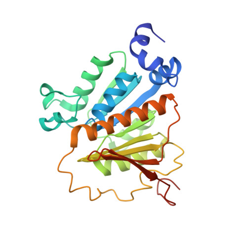
Structural Analysis of Escherichia Coli ThiF.

Duda, D.M.Walden, H.Sfondouris, J.Schulman, B.A.

(2005) J Mol Biol 349: 774-786

  • DOI: https://doi.org/10.1016/j.jmb.2005.04.011
  • Primary Citation of Related Structures:  
    1ZFN, 1ZKM

  • PubMed Abstract: 

    Escherichia coli ThiF is an enzyme in the biosynthetic cascade for generating the essential cofactor thiamin pyrophosphate. In this cascade, ThiF catalyzes adenylation of the C terminus of ThiS. We report here the crystal structures of ThiF, alone and in complex with ATP. The structures provide insight into a preference for ATP during adenylation of the protein ThiS. Additionally, the structures reveal an ordered crossover loop predicted to clamp the flexible tail of ThiS into the ThiF active site during the adenylation reaction. The importance of the crossover loop for ThiF activity is highlighted by mutational analysis. Comparison of ThiF with the structural homologues MoeB, APPBP1-UBA3, and SAE1-SAE2 reveals that the ATP-binding site, including an arginine-finger, is maintained throughout evolution, and shows divergence occurring in protein substrate-binding sites and regions devoted to unique steps in the specific function of each enzyme.


  • Organizational Affiliation

    Department of Structural Biology, St. Jude Children's Research Hospital, Memphis, TN 38105, USA.


Macromolecules
Find similar proteins by:  (by identity cutoff)  |  3D Structure
Entity ID: 1
MoleculeChains Sequence LengthOrganismDetailsImage
Adenylyltransferase thiF
A, B, C, D
253Escherichia coliMutation(s): 0 
Gene Names: thiF
EC: 2.7.7 (PDB Primary Data), 2.7.7.73 (UniProt)
UniProt
Find proteins for P30138 (Escherichia coli (strain K12))
Explore P30138 
Go to UniProtKB:  P30138
Entity Groups  
Sequence Clusters30% Identity50% Identity70% Identity90% Identity95% Identity100% Identity
UniProt GroupP30138
Sequence Annotations
Expand
  • Reference Sequence
Small Molecules
Ligands 2 Unique
IDChains Name / Formula / InChI Key2D Diagram3D Interactions
ATP
Query on ATP

Download Ideal Coordinates CCD File 
F [auth A],
H [auth B],
J [auth C],
L [auth D]
ADENOSINE-5'-TRIPHOSPHATE
C10 H16 N5 O13 P3
ZKHQWZAMYRWXGA-KQYNXXCUSA-N
ZN
Query on ZN

Download Ideal Coordinates CCD File 
E [auth A],
G [auth B],
I [auth C],
K [auth D]
ZINC ION
Zn
PTFCDOFLOPIGGS-UHFFFAOYSA-N
Experimental Data & Validation

Experimental Data

  • Method: X-RAY DIFFRACTION
  • Resolution: 2.75 Å
  • R-Value Free:  0.268 (Depositor), 0.240 (DCC) 
  • R-Value Work:  0.247 (Depositor), 0.250 (DCC) 
Space Group: F 41 3 2
Unit Cell:
Length ( Å )Angle ( ˚ )
a = 360.3α = 90
b = 360.3β = 90
c = 360.3γ = 90
Software Package:
Software NamePurpose
HKL-2000data collection
SCALEPACKdata scaling
CNSrefinement
HKL-2000data reduction
CNSphasing

Structure Validation

View Full Validation Report


Ligand Structure Quality Assessment 

Created with Raphaël 2.3.0Worse 01 BetterLigand structure goodness of fit to experimental dataBest fitted ATPClick on this verticalbar to view details

Entry History 

Deposition Data

Revision History  (Full details and data files)

  • Version 1.0: 2005-06-14
    Type: Initial release
  • Version 1.1: 2008-04-30
    Changes: Version format compliance
  • Version 1.2: 2011-07-13
    Changes: Version format compliance
  • Version 1.3: 2014-11-12
    Changes: Structure summary
  • Version 1.4: 2014-11-19
    Changes: Derived calculations
  • Version 1.5: 2024-02-14
    Changes: Data collection, Database references, Derived calculations