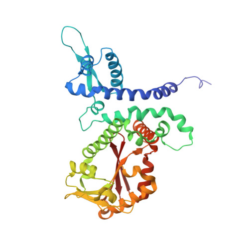
1ZG3

Crystal structure of the isoflavanone 4'-O-methyltransferase complexed with SAH and 2,7,4'-trihydroxyisoflavanone


Experimental Data Snapshot

  • Method: X-RAY DIFFRACTION
  • Resolution: 2.35 Å
  • R-Value Free: 0.266 
  • R-Value Work: 0.218 
  • R-Value Observed: 0.218 

wwPDB Validation   3D Report Full Report


Ligand Structure Quality Assessment 


This is version 1.4 of the entry. See complete history


Literature

Structural basis for dual functionality of isoflavonoid O-methyltransferases in the evolution of plant defense responses.

Liu, C.J.Deavours, B.E.Richard, S.B.Ferrer, J.L.Blount, J.W.Huhman, D.Dixon, R.A.Noel, J.P.

(2006) Plant Cell 18: 3656-3669

  • DOI: https://doi.org/10.1105/tpc.106.041376
  • Primary Citation of Related Structures:  
    1ZG3, 1ZGA, 1ZGJ, 1ZHF

  • PubMed Abstract: 

    In leguminous plants such as pea (Pisum sativum), alfalfa (Medicago sativa), barrel medic (Medicago truncatula), and chickpea (Cicer arietinum), 4'-O-methylation of isoflavonoid natural products occurs early in the biosynthesis of defense chemicals known as phytoalexins. However, among these four species, only pea catalyzes 3-O-methylation that converts the pterocarpanoid isoflavonoid 6a-hydroxymaackiain to pisatin. In pea, pisatin is important for chemical resistance to the pathogenic fungus Nectria hematococca. While barrel medic does not biosynthesize 6a-hydroxymaackiain, when cell suspension cultures are fed 6a-hydroxymaackiain, they accumulate pisatin. In vitro, hydroxyisoflavanone 4'-O-methyltransferase (HI4'OMT) from barrel medic exhibits nearly identical steady state kinetic parameters for the 4'-O-methylation of the isoflavonoid intermediate 2,7,4'-trihydroxyisoflavanone and for the 3-O-methylation of the 6a-hydroxymaackiain isoflavonoid-derived pterocarpanoid intermediate found in pea. Protein x-ray crystal structures of HI4'OMT substrate complexes revealed identically bound conformations for the 2S,3R-stereoisomer of 2,7,4'-trihydroxyisoflavanone and the 6aR,11aR-stereoisomer of 6a-hydroxymaackiain. These results suggest how similar conformations intrinsic to seemingly distinct chemical substrates allowed leguminous plants to use homologous enzymes for two different biosynthetic reactions. The three-dimensional similarity of natural small molecules represents one explanation for how plants may rapidly recruit enzymes for new biosynthetic reactions in response to changing physiological and ecological pressures.


  • Organizational Affiliation

    Howard Hughes Medical Institute, Jack H. Skirball Center for Chemical Biology and Proteomics, Salk Institute for Biological Studies, La Jolla, California, 92037, USA.


Macromolecules
Find similar proteins by:  (by identity cutoff)  |  3D Structure
Entity ID: 1
MoleculeChains Sequence LengthOrganismDetailsImage
isoflavanone 4'-O-methyltransferase358Medicago truncatulaMutation(s): 0 
Gene Names: OMT
EC: 2.1.1.46 (UniProt), 2.1.1.212 (UniProt)
UniProt
Find proteins for Q29U70 (Medicago truncatula)
Explore Q29U70 
Go to UniProtKB:  Q29U70
Entity Groups  
Sequence Clusters30% Identity50% Identity70% Identity90% Identity95% Identity100% Identity
UniProt GroupQ29U70
Sequence Annotations
Expand
  • Reference Sequence
Small Molecules
Ligands 2 Unique
IDChains Name / Formula / InChI Key2D Diagram3D Interactions
SAH
Query on SAH

Download Ideal Coordinates CCD File 
C [auth A]S-ADENOSYL-L-HOMOCYSTEINE
C14 H20 N6 O5 S
ZJUKTBDSGOFHSH-WFMPWKQPSA-N
2HI
Query on 2HI

Download Ideal Coordinates CCD File 
B [auth A](2S,3R)-2,7-DIHYDROXY-3-(4-HYDROXYPHENYL)-2,3-DIHYDRO-4H-CHROMEN-4-ONE
C15 H12 O5
YACUBWOKTPOMNW-ZFWWWQNUSA-N
Experimental Data & Validation

Experimental Data

  • Method: X-RAY DIFFRACTION
  • Resolution: 2.35 Å
  • R-Value Free: 0.266 
  • R-Value Work: 0.218 
  • R-Value Observed: 0.218 
  • Space Group: P 43 2 2
Unit Cell:
Length ( Å )Angle ( ˚ )
a = 72.106α = 90
b = 72.106β = 90
c = 188.398γ = 90
Software Package:
Software NamePurpose
CNSrefinement
HKL-2000data reduction
SCALEPACKdata scaling
CNSphasing

Structure Validation

View Full Validation Report


Ligand Structure Quality Assessment 


Entry History 

Deposition Data

Revision History  (Full details and data files)

  • Version 1.0: 2006-08-01
    Type: Initial release
  • Version 1.1: 2008-04-30
    Changes: Version format compliance
  • Version 1.2: 2011-07-13
    Changes: Derived calculations, Version format compliance
  • Version 1.3: 2014-11-12
    Changes: Structure summary
  • Version 1.4: 2024-02-14
    Changes: Data collection, Database references, Derived calculations