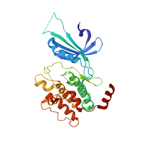
1ZY5

Crystal Structure of eIF2alpha Protein Kinase GCN2: R794G Hyperactivating Mutant Complexed with AMPPNP.


Experimental Data Snapshot

  • Method: X-RAY DIFFRACTION
  • Resolution: 2.00 Å
  • R-Value Free: 0.279 
  • R-Value Work: 0.235 

wwPDB Validation   3D Report Full Report


Ligand Structure Quality Assessment 


This is version 1.6 of the entry. See complete history


Literature

Structural Basis for Autoinhibition and Mutational Activation of Eukaryotic Initiation Factor 2{alpha} Protein Kinase GCN2

Padyana, A.K.Qiu, H.Roll-Mecak, A.Hinnebusch, A.G.Burley, S.K.

(2005) J Biol Chem 280: 29289-29299

  • DOI: https://doi.org/10.1074/jbc.M504096200
  • Primary Citation of Related Structures:  
    1ZXE, 1ZY4, 1ZY5, 1ZYC, 1ZYD

  • PubMed Abstract: 

    The GCN2 protein kinase coordinates protein synthesis with levels of amino acid stores by phosphorylating eukaryotic translation initiation factor 2. The autoinhibited form of GCN2 is activated in cells starved of amino acids by binding of uncharged tRNA to a histidyl-tRNA synthetase-like domain. Replacement of Arg-794 with Gly in the PK domain (R794G) activates GCN2 independently of tRNA binding. Crystal structures of the GCN2 protein kinase domain have been determined for wild-type and R794G mutant forms in the apo state and bound to ATP/AMPPNP. These structures reveal that GCN2 autoinhibition results from stabilization of a closed conformation that restricts ATP binding. The R794G mutant shows increased flexibility in the hinge region connecting the N- and C-lobes, resulting from loss of multiple interactions involving Arg794. This conformational change is associated with intradomain movement that enhances ATP binding and hydrolysis. We propose that intramolecular interactions following tRNA binding remodel the hinge region in a manner similar to the mechanism of enzyme activation elicited by the R794G mutation.


  • Organizational Affiliation

    Structural GenomiX, Inc., San Diego, CA 92121, USA.


Macromolecules
Find similar proteins by:  (by identity cutoff)  |  3D Structure
Entity ID: 1
MoleculeChains Sequence LengthOrganismDetailsImage
Serine/threonine-protein kinase GCN2
A, B
303Saccharomyces cerevisiaeMutation(s): 1 
Gene Names: GCN2AAS1
EC: 2.7.1.37 (PDB Primary Data), 2.7.11.1 (UniProt)
UniProt
Find proteins for P15442 (Saccharomyces cerevisiae (strain ATCC 204508 / S288c))
Explore P15442 
Go to UniProtKB:  P15442
Entity Groups  
Sequence Clusters30% Identity50% Identity70% Identity90% Identity95% Identity100% Identity
UniProt GroupP15442
Sequence Annotations
Expand
  • Reference Sequence
Experimental Data & Validation

Experimental Data

  • Method: X-RAY DIFFRACTION
  • Resolution: 2.00 Å
  • R-Value Free: 0.279 
  • R-Value Work: 0.235 
  • Space Group: P 21 21 21
Unit Cell:
Length ( Å )Angle ( ˚ )
a = 52.61α = 90
b = 78.919β = 90
c = 146.299γ = 90
Software Package:
Software NamePurpose
CNXrefinement
DENZOdata reduction
SCALEPACKdata scaling
CCP4phasing
REFMACrefinement

Structure Validation

View Full Validation Report


Ligand Structure Quality Assessment 


Entry History 

Deposition Data

Revision History  (Full details and data files)

  • Version 1.0: 2005-06-21
    Type: Initial release
  • Version 1.1: 2008-04-30
    Changes: Version format compliance
  • Version 1.2: 2011-07-13
    Changes: Version format compliance
  • Version 1.3: 2017-08-02
    Changes: Advisory, Source and taxonomy
  • Version 1.4: 2021-02-03
    Changes: Database references, Derived calculations, Structure summary
  • Version 1.5: 2021-10-20
    Changes: Advisory, Database references
  • Version 1.6: 2024-02-14
    Changes: Data collection