1ZYQ

T7 DNA polymerase in complex with 8oG and incoming ddATP


Experimental Data Snapshot

  • Method: X-RAY DIFFRACTION
  • Resolution: 2.70 Å
  • R-Value Free: 0.272 
  • R-Value Work: 0.224 
  • R-Value Observed: 0.224 

wwPDB Validation   3D Report Full Report


Ligand Structure Quality Assessment 


This is version 1.4 of the entry. See complete history


Literature

A lysine residue in the fingers subdomain of t7 DNA polymerase modulates the miscoding potential of 8-oxo-7,8-dihydroguanosine.

Brieba, L.G.Kokoska, R.J.Bebenek, K.Kunkel, T.A.Ellenberger, T.

(2005) Structure 13: 1653-1659

  • DOI: https://doi.org/10.1016/j.str.2005.07.020
  • Primary Citation of Related Structures:  
    1ZYQ

  • PubMed Abstract: 

    8-oxo-7,8-dihydroguanosine (8oG) is a highly mutagenic DNA lesion that stably pairs with adenosine, forming 8oG(syn).dA(anti) Hoogsteen base pairs. DNA polymerases show different propensities to insert dCMP or dAMP opposite 8oG, but the molecular mechanisms that determine faithful or mutagenic bypass are poorly understood. Here, we report kinetic and structural data providing evidence that, in T7 DNA polymerase, residue Lys536 is responsible for attenuating the miscoding potential of 8oG. The Lys536Ala polymerase shows a significant increase in mutagenic 8oG bypass versus wild-type polymerase, and a crystal structure of the Lys536Ala mutant reveals a closed complex with an 8oG(syn).dATP mismatch in the polymerase active site, in contrast to the unproductive, open complex previously obtained by using wild-type polymerase. We propose that Lys536 acts as a steric and/or electrostatic filter that attenuates the miscoding potential of 8oG by normally interfering with the binding of 8oG in a syn conformation that pairs with dATP.


  • Organizational Affiliation

    Department of Biological Chemistry and Molecular Pharmacology, Harvard Medical School, Massachusetts 02115, USA.


Macromolecules

Find similar proteins by:  (by identity cutoff)  |  3D Structure
Entity ID: 3
MoleculeChains Sequence LengthOrganismDetailsImage
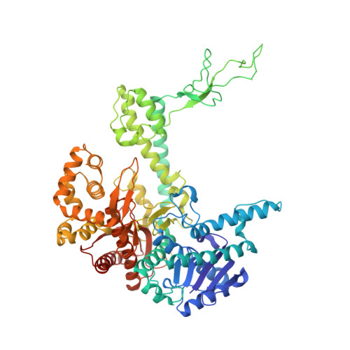
DNA polymeraseC [auth A]698Escherichia phage T7Mutation(s): 1 
Gene Names: 5
EC: 2.7.7.7 (PDB Primary Data), 3.1.11 (UniProt)
UniProt
Find proteins for P00581 (Escherichia phage T7)
Explore P00581 
Go to UniProtKB:  P00581
Entity Groups  
Sequence Clusters30% Identity50% Identity70% Identity90% Identity95% Identity100% Identity
UniProt GroupP00581
Sequence Annotations
Expand
  • Reference Sequence
Find similar proteins by:  (by identity cutoff)  |  3D Structure
Entity ID: 4
MoleculeChains Sequence LengthOrganismDetailsImage
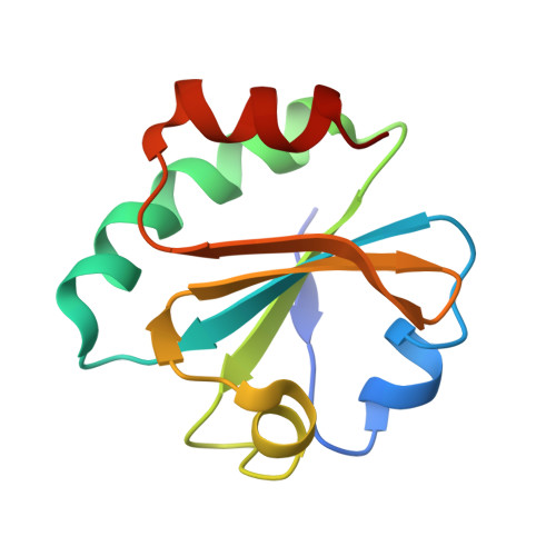
Thioredoxin 1D [auth B]108Escherichia coliMutation(s): 0 
Gene Names: trxAfipAtsnC
UniProt
Find proteins for P0AA25 (Escherichia coli (strain K12))
Explore P0AA25 
Go to UniProtKB:  P0AA25
Entity Groups  
Sequence Clusters30% Identity50% Identity70% Identity90% Identity95% Identity100% Identity
UniProt GroupP0AA25
Sequence Annotations
Expand
  • Reference Sequence

Find similar nucleic acids by:  Sequence   |   3D Structure  

Entity ID: 1
MoleculeChains LengthOrganismImage
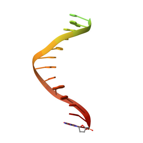
5'-D(*CP*GP*AP*AP*AP*AP*CP*GP*AP*CP*GP*GP*CP*CP*AP*GP*TP*GP*CP*CP*AP*(DDG))-3'A [auth P]22N/A
Sequence Annotations
Expand
  • Reference Sequence
Find similar nucleic acids by:  (by identity cutoff)  |  3D Structure
Entity ID: 2
MoleculeChains LengthOrganismImage
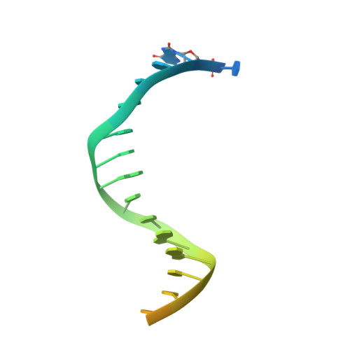
5'-D(*CP*CP*CP*(8OG)P*CP*TP*GP*GP*CP*AP*CP*TP*GP*GP*CP*CP*GP*TP*CP*GP*TP*TP*TP*TP*CP*G)-3'B [auth T]26N/A
Sequence Annotations
Expand
  • Reference Sequence
Experimental Data & Validation

Experimental Data

  • Method: X-RAY DIFFRACTION
  • Resolution: 2.70 Å
  • R-Value Free: 0.272 
  • R-Value Work: 0.224 
  • R-Value Observed: 0.224 
  • Space Group: P 21 21 2
Unit Cell:
Length ( Å )Angle ( ˚ )
a = 106.148α = 90
b = 213.007β = 90
c = 52.99γ = 90
Software Package:
Software NamePurpose
CNSrefinement
HKL-2000data reduction
SCALEPACKdata scaling
CNSphasing

Structure Validation

View Full Validation Report


Ligand Structure Quality Assessment 


Entry History 

Deposition Data

Revision History  (Full details and data files)

  • Version 1.0: 2005-11-22
    Type: Initial release
  • Version 1.1: 2008-04-30
    Changes: Version format compliance
  • Version 1.2: 2011-07-13
    Changes: Version format compliance
  • Version 1.3: 2021-10-20
    Changes: Database references, Derived calculations
  • Version 1.4: 2024-02-14
    Changes: Data collection