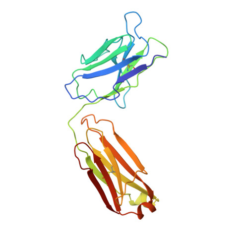
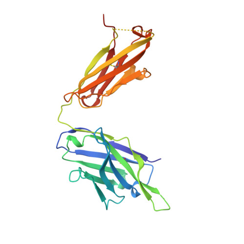
1MF2

ANTI HIV1 PROTEASE FAB COMPLEX


Experimental Data Snapshot

  • Method: X-RAY DIFFRACTION
  • Resolution: 2.60 Å
  • R-Value Free: 0.307 
  • R-Value Work: 0.206 
  • R-Value Observed: 0.206 

Starting Model: experimental
View more details

wwPDB Validation   3D Report Full Report


This is version 1.4 of the entry. See complete history


Literature

Three-dimensional structure of an Fab-peptide complex: structural basis of HIV-1 protease inhibition by a monoclonal antibody.

Lescar, J.Stouracova, R.Riottot, M.M.Chitarra, V.Brynda, J.Fabry, M.Horejsi, M.Sedlacek, J.Bentley, G.A.

(1997) J Mol Biol 267: 1207-1222

  • DOI: https://doi.org/10.1006/jmbi.1997.0950
  • Primary Citation of Related Structures:  
    1MF2, 2HRP

  • PubMed Abstract: 

    F11.2.32, a monoclonal antibody raised against HIV-1 protease (Kd = 5 nM), which inhibits proteolytic activity of the enzyme (K(inh) = 35(+/-3)nM), has been studied by crystallographic methods. The three-dimensional structure of the complex between the Fab fragment and a synthetic peptide, spanning residues 36 to 46 of the protease, has been determined at 2.2 A resolution, and that of the Fab in the free state has been determined at 2.6 A resolution. The refined model of the complex reveals ten well-ordered residues of the peptide (P36 to P45) bound in a hydrophobic cavity at the centre of the antigen-binding site. The peptide adopts a beta hairpin-like structure in which residues P38 to P42 form a type II beta-turn conformation. An intermolecular antiparallel beta-sheet is formed between the peptide and the CDR3-H loop of the antibody; additional polar interactions occur between main-chain atoms of the peptide and hydroxyl groups from tyrosine residues protruding from CDR1-L and CDR3-H. Three water molecules, located at the antigen-antibody interface, mediate polar interactions between the peptide and the most buried hypervariable loops, CDR3-L and CDR1-H. A comparison between the free and complexed Fab fragments shows that significant conformational changes occur in the long hypervariable regions, CDR1-L and CDR3-H, upon binding the peptide. The conformation of the bound peptide, which shows no overall structural similarity to the corresponding segment in HIV-1 protease, suggests that F11.2.32 might inhibit proteolysis by distorting the native structure of the enzyme.


  • Organizational Affiliation

    Unité d'Immunologie Structurale (URA 1961 CNRS), Département d'Immunologie, Institut Pasteur, Paris, France.


Macromolecules
Find similar proteins by:  (by identity cutoff)  |  3D Structure
Entity ID: 1
MoleculeChains Sequence LengthOrganismDetailsImage
MONOCLONAL ANTIBODY F11.2.32A [auth L],
C [auth M]
215Mus musculusMutation(s): 0 
UniProt
Find proteins for P01837 (Mus musculus)
Explore P01837 
Go to UniProtKB:  P01837
Entity Groups  
Sequence Clusters30% Identity50% Identity70% Identity90% Identity95% Identity100% Identity
UniProt GroupP01837
Sequence Annotations
Expand
  • Reference Sequence
Find similar proteins by:  (by identity cutoff)  |  3D Structure
Entity ID: 2
MoleculeChains Sequence LengthOrganismDetailsImage
MONOCLONAL ANTIBODY F11.2.32B [auth H],
D [auth N]
226Mus musculusMutation(s): 0 
UniProt
Find proteins for P01868 (Mus musculus)
Explore P01868 
Go to UniProtKB:  P01868
Entity Groups  
Sequence Clusters30% Identity50% Identity70% Identity90% Identity95% Identity100% Identity
UniProt GroupP01868
Sequence Annotations
Expand
  • Reference Sequence
Experimental Data & Validation

Experimental Data

  • Method: X-RAY DIFFRACTION
  • Resolution: 2.60 Å
  • R-Value Free: 0.307 
  • R-Value Work: 0.206 
  • R-Value Observed: 0.206 
  • Space Group: P 1 21 1
Unit Cell:
Length ( Å )Angle ( ˚ )
a = 67.6α = 90
b = 94.7β = 105.8
c = 70.3γ = 90
Software Package:
Software NamePurpose
DENZOdata reduction
SCALEPACKdata scaling
X-PLORmodel building
X-PLORrefinement
X-PLORphasing

Structure Validation

View Full Validation Report


Entry History 

Deposition Data

Revision History  (Full details and data files)

  • Version 1.0: 1997-12-31
    Type: Initial release
  • Version 1.1: 2008-03-24
    Changes: Version format compliance
  • Version 1.2: 2011-07-13
    Changes: Derived calculations, Version format compliance
  • Version 1.3: 2023-08-09
    Changes: Database references, Refinement description
  • Version 1.4: 2024-11-06
    Changes: Data collection, Structure summary