23KA | pdb_000023ka

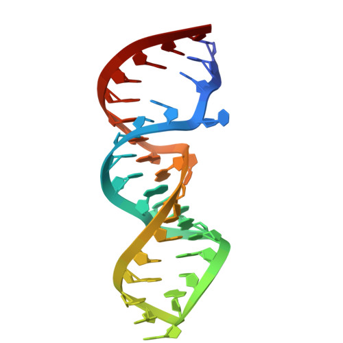
pre-miR-6074 internal loop in complex with amiloride (Form 2)


Experimental Data Snapshot

  • Method: X-RAY DIFFRACTION
  • Resolution: 2.59 Å
  • R-Value Free: 
    0.219 (Depositor), 0.221 (DCC) 
  • R-Value Work: 
    0.182 (Depositor), 0.182 (DCC) 
  • R-Value Observed: 
    0.186 (Depositor) 

Starting Model: experimental
View more details

wwPDB Validation 3D Report Full Report

Validation slider image for 23KA

Ligand Structure Quality Assessment 


This is version 1.1 of the entry. See complete history

Literature

RNA-binding landscape of amiloride: large-scale profiling and structural basis of U-U mismatch recognition.

Tsuzuki, K.Onizuka, K.Okada, M.Nagasawa, R.Miyashita, E.Komatsu, K.R.Saito, H.Kondo, J.Nagatsugi, F.

(2026) RSC Chem Biol 

  • DOI: https://doi.org/10.1039/d6cb00055j
  • Primary Citation Related Structures: 
    23JY, 23JZ, 23KA

  • PubMed Abstract: 

    Amiloride possesses a characteristic chemical scaffold capable of recognizing uracil (U) through three complementary hydrogen bonds; however, its binding selectivity toward naturally occurring RNA structural motifs has remained uncharacterized. In this study, we present a large-scale analysis of amiloride's RNA binding properties and structural characterization of the amiloride-RNA complex. Using folded RNA element profiling with structure library (FOREST), we evaluated the RNA-binding selectivity of amiloride across 3000 structured RNA motifs and uncovered pronounced binding preferences for G-quadruplexes and, notably, for a specific internal loop motif containing a U-U mismatch ( K Dapp = 0.31 µM). Furthermore, a motif extraction strategy was used to enable detailed structural investigation. The X-ray crystal structure of the amiloride-RNA complex provides the first structural evidence that amiloride recognizes a U residue within a naturally occurring RNA context via its signature complementary hydrogen bonding interactions.


  • Organizational Affiliation
    • Institute of Multidisciplinary Research for Advanced Materials, Tohoku University Miyagi 980-8577 Japan onizuka@tohoku.ac.jp nagatugi@tohoku.ac.jp.

Macromolecule Content 

  • Total Structure Weight: 34.75 kDa 
  • Atom Count: 2,333 
  • Modeled Residue Count: 105 
  • Deposited Residue Count: 105 
  • Unique nucleic acid chains: 1

Macromolecules

Find similar nucleic acids by:  (by identity cutoff) 
Entity ID: 1
MoleculeChains LengthOrganismImage
RNA (35-MER)
A, B, C
35synthetic construct
Sequence Annotations
Expand
Reference Sequence

Small Molecules

Ligands 3 Unique
IDChains Name / Formula / InChI Key2D Diagram3D Interactions
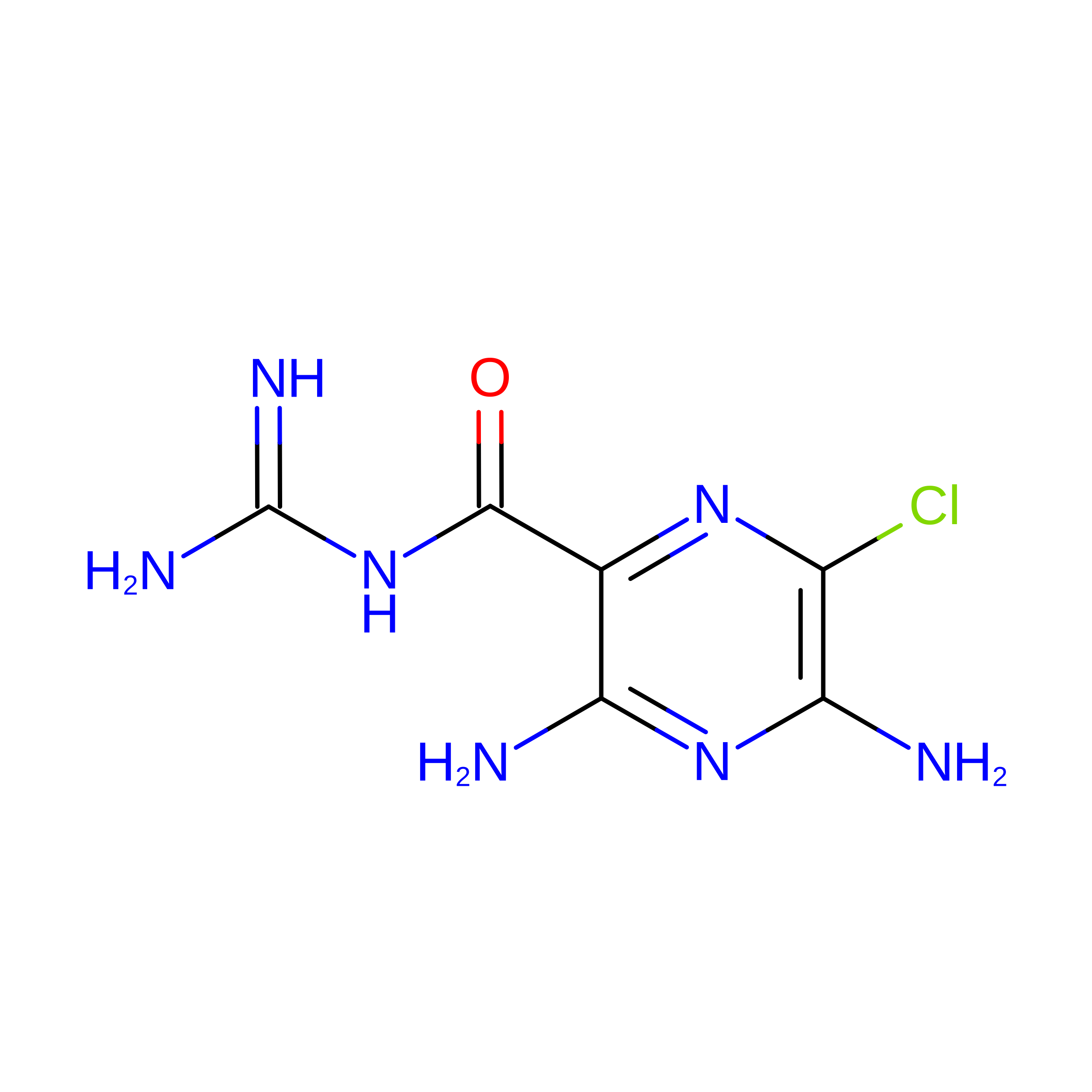
AMR
(Subject of Investigation/LOI)

Query on AMR


Download:Ideal Coordinates CCD File
D [auth A],
F [auth B],
I [auth C]
3,5-DIAMINO-N-(AMINOIMINOMETHYL)-6-CHLOROPYRAZINECARBOXAMIDE
C6 H8 Cl N7 O
XSDQTOBWRPYKKA-UHFFFAOYSA-N
CAD

Query on CAD


Download:Ideal Coordinates CCD File
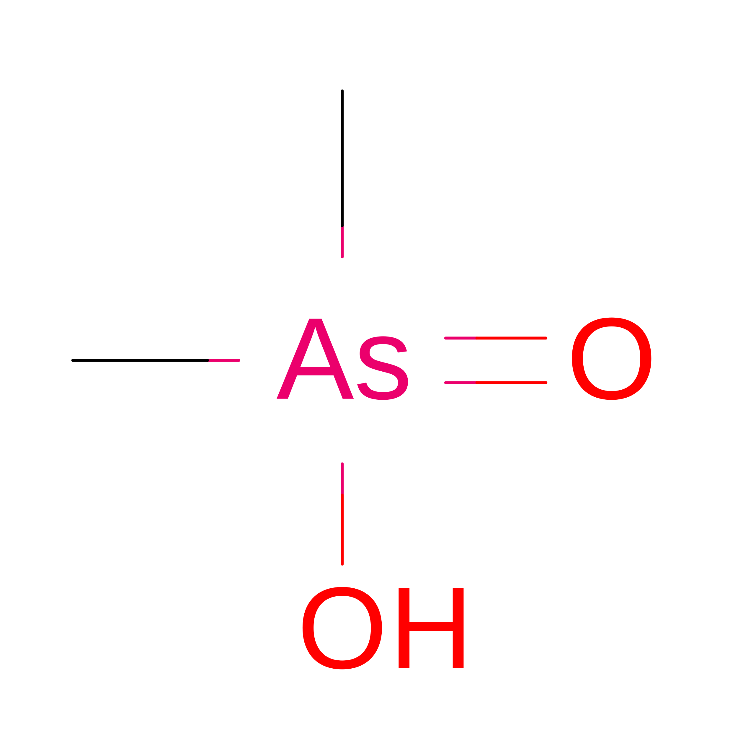
G [auth B]CACODYLIC ACID
C2 H7 As O2
OGGXGZAMXPVRFZ-UHFFFAOYSA-N
NA

Query on NA


Download:Ideal Coordinates CCD File
E [auth A],
H [auth B],
J [auth C]
SODIUM ION
Na
FKNQFGJONOIPTF-UHFFFAOYSA-N

Experimental Data & Validation

Experimental Data

  • Method: X-RAY DIFFRACTION
  • Resolution: 2.59 Å
  • R-Value Free:  0.219 (Depositor), 0.221 (DCC) 
  • R-Value Work:  0.182 (Depositor), 0.182 (DCC) 
  • R-Value Observed: 0.186 (Depositor) 
Space Group: P 32
Unit Cell:
Length ( Å )Angle ( ˚ )
a = 61.511α = 90
b = 61.511β = 90
c = 106.158γ = 120
Software Package:
Software NamePurpose
PHENIXrefinement
PDB_EXTRACTdata extraction
XDSdata reduction
XSCALEdata scaling
PHASERphasing

Structure Validation

View Full Validation Report


Ligand Structure Quality Assessment 


Entry History 

& Funding Information

Deposition Data


Funding OrganizationLocationGrant Number
Japan Agency for Medical Research and Development (AMED)JapanJP25ama121014

Revision History  (Full details and data files)

  • Version 1.0: 2026-05-06
    Type: Initial release
  • Version 1.1: 2026-05-20
    Changes: Database references