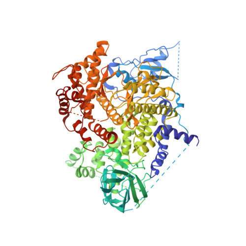
2A5U

Crystal Structure of human PI3Kgamma complexed with AS605240


Experimental Data Snapshot

  • Method: X-RAY DIFFRACTION
  • Resolution: 2.70 Å
  • R-Value Free: 0.351 
  • R-Value Work: 0.279 
  • R-Value Observed: 0.285 

Starting Model: experimental
View more details

wwPDB Validation   3D Report Full Report


Ligand Structure Quality Assessment 


This is version 1.4 of the entry. See complete history


Literature

Blockade of PI3Kgamma suppresses joint inflammation and damage in mouse models of rheumatoid arthritis

Camps, M.Ruckle, T.Ji, H.Ardissone, V.Rintelen, F.Shaw, J.Ferrandi, C.Chabert, C.Gillieron, C.Francon, B.Martin, T.Gretener, D.Perrin, D.Leroy, D.Vitte, P.-A.Hirsch, E.Wymann, M.P.Cirillo, R.Schwarz, M.K.Rommel, C.

(2005) Nat Med 11: 936-943

  • DOI: https://doi.org/10.1038/nm1284
  • Primary Citation of Related Structures:  
    2A4Z, 2A5U

  • PubMed Abstract: 

    Phosphoinositide 3-kinases (PI3K) have long been considered promising drug targets for the treatment of inflammatory and autoimmune disorders as well as cancer and cardiovascular diseases. But the lack of specificity, isoform selectivity and poor biopharmaceutical profile of PI3K inhibitors have so far hampered rigorous disease-relevant target validation. Here we describe the identification and development of specific, selective and orally active small-molecule inhibitors of PI3Kgamma (encoded by Pik3cg). We show that Pik3cg(-/-) mice are largely protected in mouse models of rheumatoid arthritis; this protection correlates with defective neutrophil migration, further validating PI3Kgamma as a therapeutic target. We also describe that oral treatment with a PI3Kgamma inhibitor suppresses the progression of joint inflammation and damage in two distinct mouse models of rheumatoid arthritis, reproducing the protective effects shown by Pik3cg(-/-) mice. Our results identify selective PI3Kgamma inhibitors as potential therapeutic molecules for the treatment of chronic inflammatory disorders such as rheumatoid arthritis.


  • Organizational Affiliation

    Serono Pharmaceutical Research Institute, Serono International S.A., 14, Chemin des Aulx, 1228 Plan-les-Ouates, Geneva, Switzerland.


Macromolecules
Find similar proteins by:  (by identity cutoff)  |  3D Structure
Entity ID: 1
MoleculeChains Sequence LengthOrganismDetailsImage
Phosphatidylinositol-4,5-bisphosphate 3-kinase catalytic subunit, gamma isoform966Homo sapiensMutation(s): 0 
EC: 2.7.1.153 (PDB Primary Data), 2.7.1.137 (UniProt), 2.7.1.154 (UniProt), 2.7.11.1 (UniProt)
UniProt & NIH Common Fund Data Resources
Find proteins for P48736 (Homo sapiens)
Explore P48736 
Go to UniProtKB:  P48736
PHAROS:  P48736
GTEx:  ENSG00000105851 
Entity Groups  
Sequence Clusters30% Identity50% Identity70% Identity90% Identity95% Identity100% Identity
UniProt GroupP48736
Sequence Annotations
Expand
  • Reference Sequence
Small Molecules
Ligands 1 Unique
IDChains Name / Formula / InChI Key2D Diagram3D Interactions
QYT
Query on QYT

Download Ideal Coordinates CCD File 
B [auth A](5E)-5-(QUINOXALIN-6-YLMETHYLENE)-1,3-THIAZOLIDINE-2,4-DIONE
C12 H7 N3 O2 S
SQWZFLMPDUSYGV-POHAHGRESA-N
Binding Affinity Annotations 
IDSourceBinding Affinity
QYT PDBBind:  2A5U IC50: 8 (nM) from 1 assay(s)
BindingDB:  2A5U IC50: min: 8, max: 9 (nM) from 2 assay(s)
Experimental Data & Validation

Experimental Data

  • Method: X-RAY DIFFRACTION
  • Resolution: 2.70 Å
  • R-Value Free: 0.351 
  • R-Value Work: 0.279 
  • R-Value Observed: 0.285 
  • Space Group: C 1 2 1
Unit Cell:
Length ( Å )Angle ( ˚ )
a = 140.446α = 90
b = 67.406β = 96.35
c = 105.971γ = 90
Software Package:
Software NamePurpose
SCALAdata scaling
CNSrefinement
PDB_EXTRACTdata extraction
MOSFLMdata reduction
CCP4data scaling
AMoREphasing
CNXrefinement

Structure Validation

View Full Validation Report


Ligand Structure Quality Assessment 


Entry History 

Revision History  (Full details and data files)

  • Version 1.0: 2005-09-20
    Type: Initial release
  • Version 1.1: 2008-04-30
    Changes: Version format compliance
  • Version 1.2: 2011-07-13
    Changes: Version format compliance
  • Version 1.3: 2017-10-11
    Changes: Refinement description
  • Version 1.4: 2023-10-25
    Changes: Data collection, Database references, Derived calculations, Refinement description