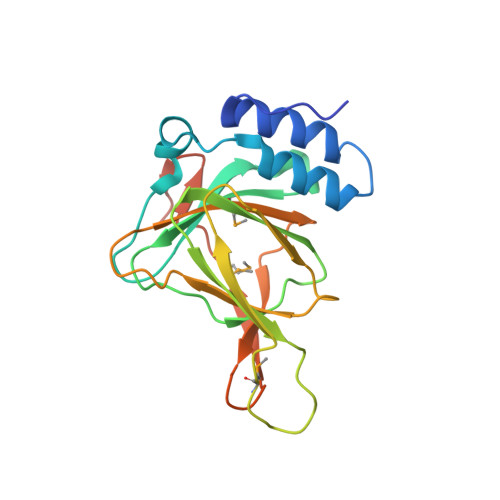
Structure and mechanism of mouse cysteine dioxygenase.
McCoy, J.G., Bailey, L.J., Bitto, E., Bingman, C.A., Aceti, D.J., Fox, B.G., Phillips, G.N.(2006) Proc Natl Acad Sci U S A 103: 3084-3089
- PubMed: 16492780
- DOI: https://doi.org/10.1073/pnas.0509262103
- Primary Citation of Related Structures:
2ATF - PubMed Abstract:
Cysteine dioxygenase (CDO) catalyzes the oxidation of l-cysteine to cysteine sulfinic acid. Deficiencies in this enzyme have been linked to autoimmune diseases and neurological disorders. The x-ray crystal structure of CDO from Mus musculus was solved to a nominal resolution of 1.75 Angstroms. The sequence is 91% identical to that of a human homolog. The structure reveals that CDO adopts the typical beta-barrel fold of the cupin superfamily. The NE2 atoms of His-86, -88, and -140 provide the metal binding site. The structure further revealed a covalent linkage between the side chains of Cys-93 and Tyr-157, the cysteine of which is conserved only in eukaryotic proteins. Metal analysis showed that the recombinant enzyme contained a mixture of iron, nickel, and zinc, with increased iron content associated with increased catalytic activity. Details of the predicted active site are used to present and discuss a plausible mechanism of action for the enzyme.
- Center for Eukaryotic Structural Genomics and Department of Biochemistry, University of Wisconsin, Madison, 53706-1544, USA.
Organizational Affiliation:


