2AZT | pdb_00002azt

Crystal structure of H176N mutant of human Glycine N-Methyltransferase


Experimental Data Snapshot

  • Method: X-RAY DIFFRACTION
  • Resolution: 2.70 Å
  • R-Value Free: 
    0.282 (Depositor) 
  • R-Value Work: 
    0.236 (Depositor) 
  • R-Value Observed: 
    0.239 (Depositor) 

Starting Model: experimental
View more details

wwPDB Validation 3D Report Full Report

Validation slider image for 2AZT

This is version 1.4 of the entry. See complete history

Literature

Destabilization of human glycine N-methyltransferase by H176N mutation.

Luka, Z.Pakhomova, S.Luka, Y.Newcomer, M.E.Wagner, C.

(2007) Protein Sci 16: 1957-1964

  • DOI: https://doi.org/10.1110/ps.072921507
  • Primary Citation Related Structures: 
    2AZT

  • PubMed Abstract: 

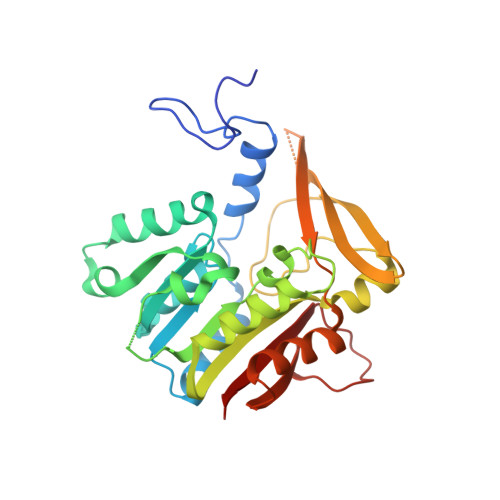
    In the presence of moderate (2-4 M) urea concentrations the tetrameric enzyme, glycine N-methyltransferase (GNMT), dissociates into compact monomers. Higher concentrations of urea (7-8 M) promote complete denaturation of the enzyme. We report here that the H176N mutation in this enzyme, found in humans with hypermethioninaemia, significantly decreases stability of the tetramer, although H176 is located far from the intersubunit contact areas. Dissociation of the tetramer to compact monomers and unfolding of compact monomers of the mutant protein were detected by circular dichroism, quenching of fluorescence emission, size-exclusion chromatography, and enzyme activity. The values of apparent free energy of dissociation of tetramer and of unfolding of compact monomers for the H176N mutant (27.7 and 4.2 kcal/mol, respectively) are lower than those of wild-type protein (37.5 and 6.2 kcal/mol). A 2.7 A resolution structure of the mutant protein revealed no significant difference in the conformation of the protein near the mutated residue.


  • Organizational Affiliation
    • Department of Biochemistry, Vanderbilt University School of Medicine, Nashville, Tennessee 37232, USA.

Macromolecule Content 

  • Total Structure Weight: 66.33 kDa 
  • Atom Count: 4,355 
  • Modeled Residue Count: 553 
  • Deposited Residue Count: 590 
  • Unique protein chains: 1

Macromolecules

Find similar proteins by:|  3D Structure
Entity ID: 1
MoleculeChains  Sequence LengthOrganismDetailsImage
Glycine N-methyltransferase
A, B
295Homo sapiensMutation(s): 1 
Gene Names: GNMT
EC: 2.1.1.20
UniProt & NIH Common Fund Data Resources
Find proteins for Q14749 (Homo sapiens)
Explore Q14749 
Go to UniProtKB:  Q14749
PHAROS:  Q14749
GTEx:  ENSG00000124713 
Entity Groups
Sequence Clusters30% Identity50% Identity70% Identity90% Identity95% Identity100% Identity
UniProt GroupQ14749
Sequence Annotations
Expand
Reference Sequence

Experimental Data & Validation

Experimental Data

  • Method: X-RAY DIFFRACTION
  • Resolution: 2.70 Å
  • R-Value Free:  0.282 (Depositor) 
  • R-Value Work:  0.236 (Depositor) 
  • R-Value Observed: 0.239 (Depositor) 
Space Group: P 21 21 2
Unit Cell:
Length ( Å )Angle ( ˚ )
a = 76.116α = 90
b = 83.279β = 90
c = 115.45γ = 90
Software Package:
Software NamePurpose
REFMACrefinement
DENZOdata reduction
SCALEPACKdata scaling
CNSphasing

Structure Validation

View Full Validation Report


Entry History 

Deposition Data

Revision History  (Full details and data files)

  • Version 1.0: 2006-09-26
    Type: Initial release
  • Version 1.1: 2007-10-16
    Changes: Version format compliance
  • Version 1.2: 2011-07-13
    Changes: Derived calculations, Version format compliance
  • Version 1.3: 2021-10-20
    Changes: Database references, Derived calculations
  • Version 1.4: 2023-08-23
    Changes: Data collection, Refinement description