2B3O

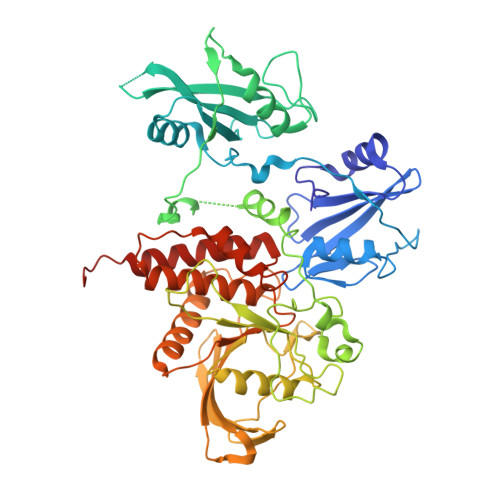
Crystal structure of human tyrosine phosphatase SHP-1


Experimental Data Snapshot

  • Method: X-RAY DIFFRACTION
  • Resolution: 2.80 Å
  • R-Value Free: 0.296 
  • R-Value Work: 0.233 
  • R-Value Observed: 0.241 

wwPDB Validation   3D Report Full Report


This is version 1.3 of the entry. See complete history


Literature

Crystal structure of human protein-tyrosine phosphatase SHP-1.

Yang, J.Liu, L.He, D.Song, X.Liang, X.Zhao, Z.J.Zhou, G.W.

(2003) J Biol Chem 278: 6516-6520

  • DOI: https://doi.org/10.1074/jbc.M210430200
  • Primary Citation of Related Structures:  
    2B3O

  • PubMed Abstract: 

    SHP-1 is a cytosolic protein-tyrosine phosphatase that behaves as a negative regulator in eukaryotic cellular signaling pathways. To understand its regulatory mechanism, we have determined the crystal structure of the C-terminal truncated human SHP-1 in the inactive conformation at 2.8-A resolution and refined the structure to a crystallographic R-factor of 24.0%. The three-dimensional structure shows that the ligand-free SHP-1 has an auto-inhibited conformation. Its N-SH2 domain blocks the catalytic domain and keeps the enzyme in the inactive conformation, which supports that the phosphatase activity of SHP-1 is primarily regulated by the N-SH2 domain. In addition, the C-SH2 domain of SHP-1 has a different orientation from and is more flexible than that of SHP-2, which enables us to propose an enzymatic activation mechanism in which the C-SH2 domains of SHPs could be involved in searching for phosphotyrosine activators.


  • Organizational Affiliation

    Program in Molecular Medicine, University of Massachusetts Medical School, Worcester, Massachusetts 01605, USA.


Macromolecules
Find similar proteins by:  (by identity cutoff)  |  3D Structure
Entity ID: 1
MoleculeChains Sequence LengthOrganismDetailsImage
Tyrosine-protein phosphatase, non-receptor type 6532Homo sapiensMutation(s): 0 
Gene Names: PTPN6HCPPTP1C
EC: 3.1.3.48
UniProt & NIH Common Fund Data Resources
Find proteins for P29350 (Homo sapiens)
Explore P29350 
Go to UniProtKB:  P29350
PHAROS:  P29350
GTEx:  ENSG00000111679 
Entity Groups  
Sequence Clusters30% Identity50% Identity70% Identity90% Identity95% Identity100% Identity
UniProt GroupP29350
Sequence Annotations
Expand
  • Reference Sequence
Experimental Data & Validation

Experimental Data

  • Method: X-RAY DIFFRACTION
  • Resolution: 2.80 Å
  • R-Value Free: 0.296 
  • R-Value Work: 0.233 
  • R-Value Observed: 0.241 
  • Space Group: P 21 21 21
Unit Cell:
Length ( Å )Angle ( ˚ )
a = 44.736α = 90
b = 100.404β = 90
c = 149.475γ = 90
Software Package:
Software NamePurpose
DENZOdata reduction
SCALAdata scaling
AMoREphasing
CNSrefinement
CCP4data scaling

Structure Validation

View Full Validation Report


Entry History 

Deposition Data

Revision History  (Full details and data files)

  • Version 1.0: 2005-10-25
    Type: Initial release
  • Version 1.1: 2008-05-01
    Changes: Version format compliance
  • Version 1.2: 2011-07-13
    Changes: Version format compliance
  • Version 1.3: 2024-02-14
    Changes: Data collection, Database references