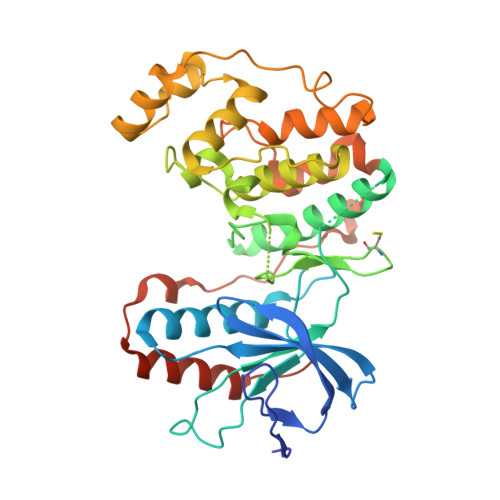
2BAQ

p38alpha bound to Ro3201195


Experimental Data Snapshot

  • Method: X-RAY DIFFRACTION
  • Resolution: 2.80 Å
  • R-Value Free: 0.296 
  • R-Value Work: 0.218 
  • R-Value Observed: 0.222 

wwPDB Validation   3D Report Full Report


Ligand Structure Quality Assessment 

Created with Raphaël 2.3.0Worse 01 BetterLigand structure goodness of fit to experimental dataBest fitted PQBClick on this verticalbar to view details

This is version 1.4 of the entry. See complete history


Literature

Prevention of MKK6-Dependent Activation by Binding to p38alpha MAP Kinase.

Sullivan, J.E.Holdgate, G.A.Campbell, D.Timms, D.Gerhardt, S.Breed, J.Breeze, A.L.Bermingham, A.Pauptit, R.A.Norman, R.A.Embrey, K.J.Read, J.Vanscyoc, W.S.Ward, W.H.

(2005) Biochemistry 44: 16475-16490

  • DOI: https://doi.org/10.1021/bi051714v
  • Primary Citation of Related Structures:  
    2BAJ, 2BAK, 2BAL, 2BAQ

  • PubMed Abstract: 

    Inhibition of p38alpha MAP kinase is a potential approach for the treatment of inflammatory disorders. MKK6-dependent phosphorylation on the activation loop of p38alpha increases its catalytic activity and affinity for ATP. An inhibitor, BIRB796, binds at a site used by the purine moiety of ATP and extends into a "selectivity pocket", which is not used by ATP. It displaces the Asp168-Phe169-Gly170 motif at the start of the activation loop, promoting a "DFG-out" conformation. Some other inhibitors bind only in the purine site, with p38alpha remaining in a "DFG-in" conformation. We now demonstrate that selectivity pocket compounds prevent MKK6-dependent activation of p38alpha in addition to inhibiting catalysis by activated p38alpha. Inhibitors using only the purine site do not prevent MKK6-dependent activation. We present kinetic analyses of seven inhibitors, whose crystal structures as complexes with p38alpha have been determined. This work includes four new crystal structures and a novel assay to measure K(d) for nonactivated p38alpha. Selectivity pocket compounds associate with p38alpha over 30-fold more slowly than purine site compounds, apparently due to low abundance of the DFG-out conformation. At concentrations that inhibit cellular production of an inflammatory cytokine, TNFalpha, selectivity pocket compounds decrease levels of phosphorylated p38alpha and beta. Stabilization of a DFG-out conformation appears to interfere with recognition of p38alpha as a substrate by MKK6. ATP competes less effectively for prevention of activation than for inhibition of catalysis. By binding to a different conformation of the enzyme, compounds that prevent activation offer an alternative approach to modulation of p38alpha.


  • Organizational Affiliation

    Molecular Enzymology, AstraZeneca, Mereside, Alderley Park, Macclesfield, Cheshire SK10 4TG, UK.


Macromolecules
Find similar proteins by:  (by identity cutoff)  |  3D Structure
Entity ID: 1
MoleculeChains Sequence LengthOrganismDetailsImage
Mitogen-activated protein kinase 14365Homo sapiensMutation(s): 2 
Gene Names: MAPK14CSBPCSBP1CSBP2MXI2
EC: 2.7.1.37 (PDB Primary Data), 2.7.11.24 (UniProt)
UniProt & NIH Common Fund Data Resources
Find proteins for Q16539 (Homo sapiens)
Explore Q16539 
Go to UniProtKB:  Q16539
PHAROS:  Q16539
GTEx:  ENSG00000112062 
Entity Groups  
Sequence Clusters30% Identity50% Identity70% Identity90% Identity95% Identity100% Identity
UniProt GroupQ16539
Sequence Annotations
Expand
  • Reference Sequence
Small Molecules
Ligands 1 Unique
IDChains Name / Formula / InChI Key2D Diagram3D Interactions
PQB
Query on PQB

Download Ideal Coordinates CCD File 
B [auth A][5-AMINO-1-(4-FLUOROPHENYL)-1H-PYRAZOL-4-YL](3-{[(2R)-2,3-DIHYDROXYPROPYL]OXY}PHENYL)METHANONE
C19 H18 F N3 O4
IJDQETGUEUJVTB-HNNXBMFYSA-N
Modified Residues  1 Unique
IDChains TypeFormula2D DiagramParent
CSS
Query on CSS
A
L-PEPTIDE LINKINGC3 H7 N O2 S2CYS
Binding Affinity Annotations 
IDSourceBinding Affinity
PQB BindingDB:  2BAQ IC50: min: 180, max: 700 (nM) from 5 assay(s)
Experimental Data & Validation

Experimental Data

  • Method: X-RAY DIFFRACTION
  • Resolution: 2.80 Å
  • R-Value Free: 0.296 
  • R-Value Work: 0.218 
  • R-Value Observed: 0.222 
  • Space Group: P 21 21 21
Unit Cell:
Length ( Å )Angle ( ˚ )
a = 64.759α = 90
b = 74.718β = 90
c = 76.929γ = 90
Software Package:
Software NamePurpose
REFMACrefinement
MOSFLMdata reduction
CCP4data scaling
AMoREphasing

Structure Validation

View Full Validation Report


Ligand Structure Quality Assessment 

Created with Raphaël 2.3.0Worse 01 BetterLigand structure goodness of fit to experimental dataBest fitted PQBClick on this verticalbar to view details

Entry History 

Deposition Data

Revision History  (Full details and data files)

  • Version 1.0: 2005-12-06
    Type: Initial release
  • Version 1.1: 2008-05-01
    Changes: Version format compliance
  • Version 1.2: 2011-07-13
    Changes: Advisory, Version format compliance
  • Version 1.3: 2021-10-20
    Changes: Database references, Derived calculations
  • Version 1.4: 2024-10-30
    Changes: Data collection, Structure summary