2C7A

STRUCTURE OF THE PROGESTERONE RECEPTOR-DNA COMPLEX


Experimental Data Snapshot

  • Method: X-RAY DIFFRACTION
  • Resolution: 2.50 Å
  • R-Value Free: 0.246 
  • R-Value Work: 0.198 
  • R-Value Observed: 0.203 

wwPDB Validation   3D Report Full Report


This is version 1.2 of the entry. See complete history


Literature

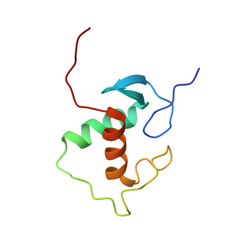
Structure of the Progesterone Receptor-Deoxyribonucleic Acid Complex: Novel Interactions Required for Binding to Half-Site Response Elements.

Roemer, S.C.Donham, D.C.Sherman, L.Pon, V.H.Edwards, D.P.Churchill, M.E.A.

(2006) Mol Endocrinol 20: 3042

  • DOI: https://doi.org/10.1210/me.2005-0511
  • Primary Citation of Related Structures:  
    2C7A

  • PubMed Abstract: 

    The DNA binding domain (DBD) of nuclear hormone receptors contains a highly conserved globular domain and a less conserved carboxyl-terminal extension (CTE). Despite previous observations that the CTEs of some classes of nuclear receptors are structured and interact with DNA outside of the hexanucleotide hormone response element (HRE), there has been no evidence for such a CTE among the steroid receptors. We have determined the structure of the progesterone receptor (PR)-DBD-CTE DNA complex at a resolution of 2.5 A, which revealed binding of the CTE to the minor groove flanking the HREs. Alanine substitutions of the interacting CTE residues reduced affinity for inverted repeat HREs separated by three nucleotides, and essentially abrogated binding to a single HRE. A highly compressed minor groove of the trinucleotide spacer and a novel dimerization interface were also observed. A PR binding site selection experiment revealed sequence preferences in the trinucleotide spacer and flanking DNA. These results, taken together, support the notion that sequences outside of the HREs influence the DNA binding affinity and specificity of steroid receptors.


  • Organizational Affiliation

    Program in Molecular Biology, Department of Pharmacology, University of Colorado at Denver and Health Sciences Center, Aurora, Colorado 80045, USA.


Macromolecules

Find similar proteins by:  (by identity cutoff)  |  3D Structure
Entity ID: 1
MoleculeChains Sequence LengthOrganismDetailsImage
PROGESTERONE RECEPTOR
A, B
78Homo sapiensMutation(s): 0 
UniProt & NIH Common Fund Data Resources
Find proteins for P06401 (Homo sapiens)
Explore P06401 
Go to UniProtKB:  P06401
PHAROS:  P06401
GTEx:  ENSG00000082175 
Entity Groups  
Sequence Clusters30% Identity50% Identity70% Identity90% Identity95% Identity100% Identity
UniProt GroupP06401
Sequence Annotations
Expand
  • Reference Sequence

Find similar nucleic acids by:  Sequence   |   3D Structure  

Entity ID: 2
MoleculeChains LengthOrganismImage
5'-D(*CP*CP*AP*GP*AP*AP*CP*AP*GP*TP *TP*TP*GP*TP*TP*CP*TP*G)-3'18Homo sapiens
Sequence Annotations
Expand
  • Reference Sequence

Find similar nucleic acids by:  Sequence   |   3D Structure  

Entity ID: 3
MoleculeChains LengthOrganismImage
5'-D(*CP*CP*AP*GP*AP*AP*CP*AP*AP*AP *CP*TP*GP*TP*TP*CP*TP*G)-3'18Homo sapiens
Sequence Annotations
Expand
  • Reference Sequence
Experimental Data & Validation

Experimental Data

  • Method: X-RAY DIFFRACTION
  • Resolution: 2.50 Å
  • R-Value Free: 0.246 
  • R-Value Work: 0.198 
  • R-Value Observed: 0.203 
  • Space Group: P 21 21 21
Unit Cell:
Length ( Å )Angle ( ˚ )
a = 39.53α = 90
b = 107.85β = 90
c = 111.96γ = 90
Software Package:
Software NamePurpose
REFMACrefinement

Structure Validation

View Full Validation Report


Entry History 

Deposition Data

Revision History  (Full details and data files)

  • Version 1.0: 2006-08-30
    Type: Initial release
  • Version 1.1: 2011-05-08
    Changes: Version format compliance
  • Version 1.2: 2011-07-13
    Changes: Version format compliance