2CG5

Structure of aminoadipate-semialdehyde dehydrogenase- phosphopantetheinyl transferase in complex with cytosolic acyl carrier protein and coenzyme A


Experimental Data Snapshot

  • Method: X-RAY DIFFRACTION
  • Resolution: 2.70 Å
  • R-Value Free: 0.247 
  • R-Value Work: 0.192 
  • R-Value Observed: 0.195 

wwPDB Validation   3D Report Full Report


Ligand Structure Quality Assessment 


This is version 1.4 of the entry. See complete history


Literature

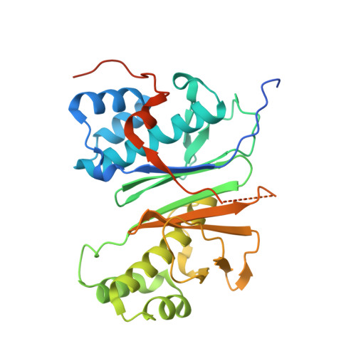
Mechanism and Substrate Recognition of Human Holo Acp Synthase.

Bunkoczi, G.Pasta, S.Joshi, A.Wu, X.Kavanagh, K.L.Smith, S.Oppermann, U.

(2007) Chem Biol 14: 1243

  • DOI: https://doi.org/10.1016/j.chembiol.2007.10.013
  • Primary Citation of Related Structures:  
    2BYD, 2C43, 2CG5

  • PubMed Abstract: 

    Mammals utilize a single phosphopantetheinyl transferase for the posttranslational modification of at least three different apoproteins: the carrier protein components of cytosolic and mitochondrial fatty acid synthases and the aminoadipate semialdehyde reductase involved in lysine degradation. We determined the crystal structure of the human phosphopantetheinyl transferase, a eukaryotic phosphopantetheinyl transferase characterized, complexed with CoA and Mg(2+), and in ternary complex with CoA and ACP. The involvement of key residues in ligand binding and catalysis was confirmed by mutagenesis and kinetic analysis. Human phosphopantetheinyl transferase exhibits an alpha/beta fold and 2-fold pseudosymmetry similar to the Sfp phosphopantetheinyl transferase from Bacillus subtilis. Although the bound ACP exhibits a typical four-helix structure, its binding is unusual in that it is facilitated predominantly by hydrophobic interactions. A detailed mechanism is proposed describing the substrate binding and catalytic process.


  • Organizational Affiliation

    Structural Genomics Consortium, University of Oxford, Oxford OX3 7LD, UK.


Macromolecules
Find similar proteins by:  (by identity cutoff)  |  3D Structure
Entity ID: 1
MoleculeChains Sequence LengthOrganismDetailsImage
L-AMINOADIPATE-SEMIALDEHYDE DEHYDROGENASE-PHOSPHOPANTETHEINYL TRANSFERASE319Homo sapiensMutation(s): 0 
EC: 1.2.1.31
UniProt & NIH Common Fund Data Resources
Find proteins for Q9NRN7 (Homo sapiens)
Explore Q9NRN7 
Go to UniProtKB:  Q9NRN7
PHAROS:  Q9NRN7
GTEx:  ENSG00000149313 
Entity Groups  
Sequence Clusters30% Identity50% Identity70% Identity90% Identity95% Identity100% Identity
UniProt GroupQ9NRN7
Sequence Annotations
Expand
  • Reference Sequence
Find similar proteins by:  (by identity cutoff)  |  3D Structure
Entity ID: 2
MoleculeChains Sequence LengthOrganismDetailsImage
FATTY ACID SYNTHASE91Homo sapiensMutation(s): 1 
EC: 2.3.1.85
UniProt & NIH Common Fund Data Resources
Find proteins for P49327 (Homo sapiens)
Explore P49327 
Go to UniProtKB:  P49327
PHAROS:  P49327
GTEx:  ENSG00000169710 
Entity Groups  
Sequence Clusters30% Identity50% Identity70% Identity90% Identity95% Identity100% Identity
UniProt GroupP49327
Sequence Annotations
Expand
  • Reference Sequence
Experimental Data & Validation

Experimental Data

  • Method: X-RAY DIFFRACTION
  • Resolution: 2.70 Å
  • R-Value Free: 0.247 
  • R-Value Work: 0.192 
  • R-Value Observed: 0.195 
  • Space Group: P 32 2 1
Unit Cell:
Length ( Å )Angle ( ˚ )
a = 69.361α = 90
b = 69.361β = 90
c = 184.715γ = 120
Software Package:
Software NamePurpose
REFMACrefinement
XDSdata reduction
XDSdata scaling
PHASERphasing

Structure Validation

View Full Validation Report


Ligand Structure Quality Assessment 


Entry History 

Deposition Data

Revision History  (Full details and data files)

  • Version 1.0: 2006-03-01
    Type: Initial release
  • Version 1.1: 2011-07-13
    Changes: Advisory, Refinement description, Version format compliance
  • Version 1.2: 2018-01-24
    Changes: Structure summary
  • Version 1.3: 2018-04-04
    Changes: Data collection
  • Version 1.4: 2023-12-13
    Changes: Data collection, Database references, Derived calculations, Other, Refinement description