2CLZ | pdb_00002clz

Mhc Class I Natural Mutant H-2Kbm8 Heavy Chain Complexed With beta-2 Microglobulin and pBM1 peptide


Experimental Data Snapshot

  • Method: X-RAY DIFFRACTION
  • Resolution: 1.90 Å
  • R-Value Free: 
    0.247 (Depositor) 
  • R-Value Work: 
    0.206 (Depositor) 
  • R-Value Observed: 
    0.208 (Depositor) 

Starting Model: experimental
View more details

wwPDB Validation 3D Report Full Report

Validation slider image for 2CLZ

This is version 1.4 of the entry. See complete history

Literature

Distinct Orientation of the Alloreactive Monoclonal Cd8 T Cell Activation Program by Three Different Peptide/Mhc Complexes.

Auphan-Anezin, N.Mazza, C.Guimezanes, A.Barrett-Wilt, G.A.Montero-Julian, F.Roussel, A.Hunt, D.F.Malissen, B.Schmitt-Verhulst, A.M.

(2006) Eur J Immunol 36: 1856

  • DOI: https://doi.org/10.1002/eji.200635895
  • Primary Citation Related Structures: 
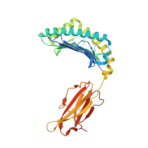
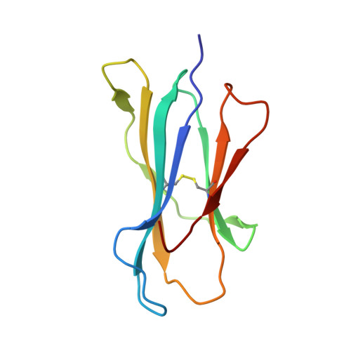
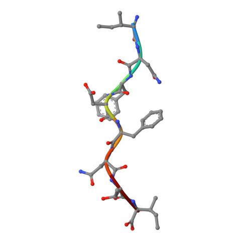
    2CLV, 2CLZ

  • PubMed Abstract: 

    We have characterized three different programs of activation for alloreactive CD8 T cells expressing the BM3.3 TCR, their elicitation depending on the characteristics of the stimulating peptide/MHC complex. The high-affinity interaction between the TCR and the K(b)-associated endogenous peptide pBM1 (INFDFNTI) induced a complete differentiation program into effector cells correlated with sustained ERK activation. The K(bm8) variant elicited a partial activation program with delayed T cell proliferation, poor CTL activity and undetectable ERK phosphorylation; this resulted from a low-avidity interaction of TCR BM3.3 with a newly identified endogenous peptide, pBM8 (SQYYYNSL). Interestingly, mismatched pBM1/K(bm8) complexes induced a split response in BM3.3 T cells, with total reconstitution of T cell proliferation but defective generation of CTL activity that was correlated with strong but shortened ERK phosphorylation. Crystal structures highlight the molecular basis for the higher stability of pBM8/K(bm8) compared to pBM1/K(bm8) complexes that exist in two conformers. This study illustrates the importance of the stability of both peptide/MHC and peptide/MHC-TCR interactions for induction of sustained signaling required to induce optimal CTL effector functions. Subtle allelic structural variations, amplified by peptide selection, may thus orient distinct outcomes of alloreactive TCR-based therapies.


  • Organizational Affiliation
    • Centre d'Immunologie de Marseille-Luminy, CNRS-INSERM-Universite de la Méditerranée, Campus de Luminy, Marseille, France. auphan@ciml.univ-mrs.fr

Macromolecule Content 

  • Total Structure Weight: 89.53 kDa 
  • Atom Count: 6,939 
  • Modeled Residue Count: 772 
  • Deposited Residue Count: 772 
  • Unique protein chains: 3

Macromolecules

Find similar proteins by:|  3D Structure
Entity ID: 1
MoleculeChains  Sequence LengthOrganismDetailsImage
H-2 CLASS I HISTOCOMPATIBILITY ANTIGEN, K-B ALPHA CHAINA,
D [auth H]
279Mus musculusMutation(s): 4 
UniProt
Find proteins for P01901 (Mus musculus)
Explore P01901 
Go to UniProtKB:  P01901
Entity Groups
Sequence Clusters30% Identity50% Identity70% Identity90% Identity95% Identity100% Identity
UniProt GroupP01901
Sequence Annotations
Expand
Reference Sequence
Find similar proteins by:|  3D Structure
Entity ID: 2
MoleculeChains  Sequence LengthOrganismDetailsImage
BETA-2 MICROGLOBULINB,
F [auth P]
99Mus musculusMutation(s): 0 
UniProt & NIH Common Fund Data Resources
Find proteins for P01887 (Mus musculus)
Explore P01887 
Go to UniProtKB:  P01887
IMPC:  MGI:88127
Entity Groups
Sequence Clusters30% Identity50% Identity70% Identity90% Identity95% Identity100% Identity
UniProt GroupP01887
Sequence Annotations
Expand
Reference Sequence
Find similar proteins by:  Sequence   |   3D Structure  
Entity ID: 3
MoleculeChains  Sequence LengthOrganismDetailsImage
RBM5 PROTEINC,
E [auth M]
8Mus musculusMutation(s): 0 
UniProt
Find proteins for Q8CDD8 (Mus musculus)
Explore Q8CDD8 
Go to UniProtKB:  Q8CDD8
Entity Groups
UniProt GroupQ8CDD8
Sequence Annotations
Expand
Reference Sequence

Experimental Data & Validation

Experimental Data

  • Method: X-RAY DIFFRACTION
  • Resolution: 1.90 Å
  • R-Value Free:  0.247 (Depositor) 
  • R-Value Work:  0.206 (Depositor) 
  • R-Value Observed: 0.208 (Depositor) 
Space Group: P 1 21 1
Unit Cell:
Length ( Å )Angle ( ˚ )
a = 66.588α = 90
b = 90.759β = 111.35
c = 89.248γ = 90
Software Package:
Software NamePurpose
REFMACrefinement
MOSFLMdata reduction
SCALAdata scaling
CNSphasing

Structure Validation

View Full Validation Report


Entry History 

Revision History  (Full details and data files)

  • Version 1.0: 2006-06-14
    Type: Initial release
  • Version 1.1: 2011-05-08
    Changes: Version format compliance
  • Version 1.2: 2011-07-13
    Changes: Version format compliance
  • Version 1.3: 2023-12-13
    Changes: Data collection, Database references, Other, Refinement description
  • Version 1.4: 2024-11-13
    Changes: Structure summary