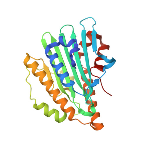
Crystal structure of phycocyanobilin:ferredoxin oxidoreductase in complex with biliverdin IXalpha, a key enzyme in the biosynthesis of phycocyanobilin
Hagiwara, Y., Sugishima, M., Takahashi, Y., Fukuyama, K.(2006) Proc Natl Acad Sci U S A 103: 27-32
- PubMed: 16380422
- DOI: https://doi.org/10.1073/pnas.0507266103
- Primary Citation of Related Structures:
2D1E - PubMed Abstract:
Phytobilins (light harvesting and photoreceptor pigments in higher plants, algae, and cyanobacteria) are synthesized from biliverdin IXalpha (BV) by ferredoxin-dependent bilin reductases (FDBRs). Phycocyanobilin:ferredoxin oxidoreductase (PcyA), one such FDBR, is a new class of radical enzymes that require neither cofactors nor metals and serially reduces the vinyl group of the D-ring and A-ring of BV using four electrons from ferredoxin to produce phycocyanobilin, one of the phytobilins. We have determined the crystal structure of PcyA from Synechocystis sp. PCC 6803 in complex with BV, revealing the first tertiary structure of an FDBR family member. PcyA is folded in a three-layer alpha/beta/alpha sandwich structure, in which BV in a cyclic conformation is positioned between the beta-sheet and C-terminal alpha-helices. The basic patch on the PcyA surface near the BV molecule may provide a binding site for acidic ferredoxin, allowing direct transfer of electrons to BV. The orientation of BV is definitely fixed in PcyA by several hydrophilic interactions and the shape of the BV binding pocket of PcyA. We propose the mechanism by which the sequential reduction of the D- and A-rings is controlled, where Asp-105, located between the two reduction sites, would play the central role by changing its conformation during the reaction. Homology modeling of other FDBRs based on the PcyA structure fits well with previous genetic and biochemical data, thereby providing a structural basis for the reaction mechanism of FDBRs.
- Department of Biology, Graduate School of Science, Osaka University, Toyonaka, Osaka 560-0043, Japan.
Organizational Affiliation:


