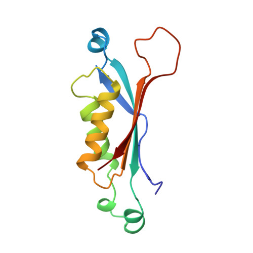
Crystal structure and reaction mechanism of 7,8-dihydroneopterin aldolase from Staphylococcus aureus.
Hennig, M., D'Arcy, A., Hampele, I.C., Page, M.G., Oefner, C., Dale, G.E.(1998) Nat Struct Biol 5: 357-362
- PubMed: 9586996
- DOI: https://doi.org/10.1038/nsb0598-357
- Primary Citation of Related Structures:
1DHN, 2DHN - PubMed Abstract:
Dihydroneopterin aldolase catalyzes the conversion of 7,8-dihydroneopterin to 6-hydroxymethyl-7,8-dihydropterin during the de novo synthesis of folic acid from guanosine triphosphate. The gene encoding the dihydroneopterin aldolase from S. aureus has been cloned, sequenced and expressed in Escherichia coli. The protein has been purified for biochemical characterization and its X-ray structure determined at 1.65 A resolution. The protein forms an octamer of 110,000 Mr molecular weight. Four molecules assemble into a ring, and two rings come together to give a cylinder with a hole of at least 13 A diameter. The structure of the binary complex with the product 6-hydroxymethyl-7,8-dihydropterin has defined the location of the active site. The structural information and results of site directed mutagenesis allow an enzyme reaction mechanism to be proposed.


