2EXM | pdb_00002exm

Human CDK2 in complex with isopentenyladenine


Experimental Data Snapshot

  • Method: X-RAY DIFFRACTION
  • Resolution: 1.80 Å
  • R-Value Free: 
    0.270 (Depositor) 
  • R-Value Work: 
    0.200 (Depositor) 

wwPDB Validation 3D Report Full Report

Validation slider image for 2EXM

This is version 1.4 of the entry. See complete history

Literature

Multiple modes of ligand recognition: crystal structures of cyclin-dependent protein kinase 2 in complex with ATP and two inhibitors, olomoucine and isopentenyladenine.

Schulze-Gahmen, U.Brandsen, J.Jones, H.D.Morgan, D.O.Meijer, L.Vesely, J.Kim, S.H.

(1995) Proteins 22: 378-391

  • DOI: https://doi.org/10.1002/prot.340220408
  • Primary Citation Related Structures: 
    1W0X, 2EXM

  • PubMed Abstract: 

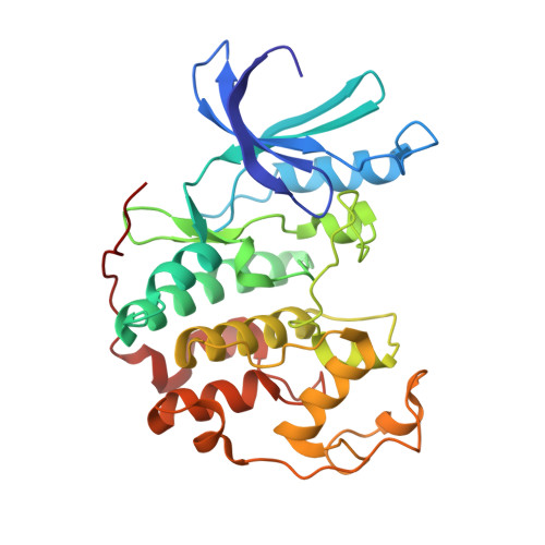
    Cyclin-dependent kinases (CDKs) are conserved regulators of the eukaryotic cell cycle with different isoforms controlling specific phases of the cell cycle. Mitogenic or growth inhibitory signals are mediated, respectively, by activation or inhibition of CDKs which phosphorylate proteins associated with the cell cycle. The central role of CDKs in cell cycle regulation makes them a potential new target for inhibitory molecules with anti-proliferative and/or anti-neoplastic effects. We describe the crystal structures of the complexes of CDK2 with a weakly specific CDK inhibitor, N6-(delta 2-isopentenyl)adenine, and a strongly specific inhibitor, olomoucine. Both inhibitors are adenine derivatives and bind in the adenine binding pocket of CDK2, but in an unexpected and different orientation from the adenine of the authentic ligand ATP. The N6-benzyl substituent in olomoucine binds outside the conserved binding pocket and is most likely responsible for its specificity. The structural information from the CDK2-olomoucine complex will be useful in directing the search for the next generation inhibitors with improved properties.


  • Organizational Affiliation
    • Department of Chemistry, University of California, Berkeley 94720, USA.

Macromolecule Content 

  • Total Structure Weight: 34.18 kDa 
  • Atom Count: 2,512 
  • Modeled Residue Count: 298 
  • Deposited Residue Count: 298 
  • Unique protein chains: 1

Macromolecules

Find similar proteins by:|  3D Structure
Entity ID: 1
MoleculeChains  Sequence LengthOrganismDetailsImage
Cell division protein kinase 2298Homo sapiensMutation(s): 0 
Gene Names: CDK2
EC: 2.7.1.37 (PDB Primary Data), 2.7.11.22 (UniProt)
UniProt & NIH Common Fund Data Resources
Find proteins for P24941 (Homo sapiens)
Explore P24941 
Go to UniProtKB:  P24941
PHAROS:  P24941
GTEx:  ENSG00000123374 
Entity Groups
Sequence Clusters30% Identity50% Identity70% Identity90% Identity95% Identity100% Identity
UniProt GroupP24941
Sequence Annotations
Expand
Reference Sequence

Small Molecules

Ligands 1 Unique
IDChains Name / Formula / InChI Key2D Diagram3D Interactions
ZIP

Query on ZIP


Download:Ideal Coordinates CCD File
B [auth A]N-(3-METHYLBUT-2-EN-1-YL)-9H-PURIN-6-AMINE
C10 H13 N5
HYVABZIGRDEKCD-UHFFFAOYSA-N

Experimental Data & Validation

Experimental Data

  • Method: X-RAY DIFFRACTION
  • Resolution: 1.80 Å
  • R-Value Free:  0.270 (Depositor) 
  • R-Value Work:  0.200 (Depositor) 
Space Group: P 21 21 21
Unit Cell:
Length ( Å )Angle ( ˚ )
a = 73.25α = 90
b = 72.51β = 90
c = 54.03γ = 90
Software Package:
Software NamePurpose
X-PLORmodel building
X-PLORrefinement
X-PLORphasing

Structure Validation

View Full Validation Report


Entry History 

Deposition Data

Revision History  (Full details and data files)

  • Version 1.0: 2005-12-27
    Type: Initial release
  • Version 1.1: 2008-05-01
    Changes: Version format compliance
  • Version 1.2: 2011-07-13
    Changes: Version format compliance
  • Version 1.3: 2017-10-18
    Changes: Refinement description
  • Version 1.4: 2024-02-14
    Changes: Data collection, Database references, Derived calculations