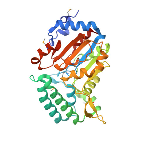
Structure of pyrimidine 5'-nucleotidase type 1. Insight into mechanism of action and inhibition during lead poisoning.
Bitto, E., Bingman, C.A., Wesenberg, G.E., McCoy, J.G., Phillips, G.N.(2006) J Biological Chem 281: 20521-20529
- PubMed: 16672222
- DOI: https://doi.org/10.1074/jbc.M602000200
- Primary Citation of Related Structures:
2BDU, 2G06, 2G07, 2G08, 2G09, 2G0A - PubMed Abstract:
Eukaryotic pyrimidine 5'-nucleotidase type 1 (P5N-1) catalyzes dephosphorylation of pyrimidine 5'-mononucleotides. Deficiency of P5N-1 activity in red blood cells results in nonspherocytic hemolytic anemia. The enzyme deficiency is either familial or can be acquired through lead poisoning. We present the crystal structure of mouse P5N-1 refined to 2.35 A resolution. The mouse P5N-1 has a 92% sequence identity to its human counterpart. The structure revealed that P5N-1 adopts a fold similar to enzymes of the haloacid dehydrogenase superfamily. The active site of this enzyme is structurally highly similar to those of phosphoserine phosphatases. We propose a catalytic mechanism for P5N-1 that is also similar to that of phosphoserine phosphatases and provide experimental evidence for the mechanism in the form of structures of several reaction cycle states, including: 1) P5N-1 with bound Mg(II) at 2.25 A, 2) phosphoenzyme intermediate analog at 2.30 A, 3) product-transition complex analog at 2.35 A, and 4) product complex at 2.1A resolution with phosphate bound in the active site. Furthermore the structure of Pb(II)-inhibited P5N-1 (at 2.35 A) revealed that Pb(II) binds within the active site in a way that compromises function of the cationic cavity, which is required for the recognition and binding of the phosphate group of nucleotides.
- Center for Eukaryotic Structural Genomics, University of Wisconsin, Madison, Wisconsin 53706-1544, USA.
Organizational Affiliation:


