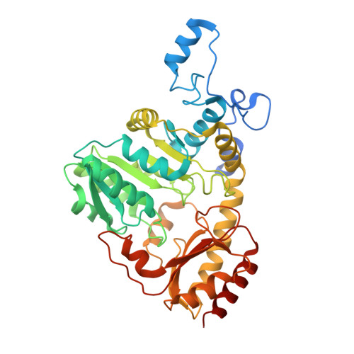
2GQN

Cystathionine Beta-Lyase (CBL) from Escherichia Coli in complex with N-Hydrazinocarbonylmethyl-2-Nitro-Benzamide


Experimental Data Snapshot

  • Method: X-RAY DIFFRACTION
  • Resolution: 1.80 Å
  • R-Value Free: 0.227 
  • R-Value Work: 0.174 
  • R-Value Observed: 0.177 

wwPDB Validation   3D Report Full Report


Ligand Structure Quality Assessment 


This is version 1.3 of the entry. See complete history


Literature

Inhibitors of Bacterial Cystathionine beta-Lyase: Leads for New Antimicrobial Agents and Probes of Enzyme Structure and Function.

Ejim, L.J.Blanchard, J.E.Koteva, K.P.Sumerfield, R.Elowe, N.H.Chechetto, J.D.Brown, E.D.Junop, M.S.Wright, G.D.

(2007) J Med Chem 50: 755-764

  • DOI: https://doi.org/10.1021/jm061132r
  • Primary Citation of Related Structures:  
    2FQ6, 2GQN

  • PubMed Abstract: 

    The biosynthesis of methionine is an attractive antibiotic target given its importance in protein and DNA metabolism and its absence in mammals. We have performed a high-throughput screen of the methionine biosynthesis enzyme cystathionine beta-lyase (CBL) against a library of 50 000 small molecules and have identified several compounds that inhibit CBL enzyme activity in vitro. These hit molecules were of two classes: those that blocked CBL activity with mixed steady-state inhibition and those that covalently interacted with the enzyme at the active site pyridoxal phosphate cofactor with slow-binding inhibition kinetics. We determined the crystal structure of one of the slow-binding inhibitors in complex with CBL and used this structure as a guide in the synthesis of a small, focused library of analogues, some of which had improved enzyme inhibition properties. These studies provide the first lead molecules for antimicrobial agents that target cystathionine beta-lyase in methionine biosynthesis.


  • Organizational Affiliation

    Antimicrobial Research Centre, McMaster High Throughput Screening Laboratory, Department of Biochemistry and Biomedical Sciences, McMaster University, Ontario L8N 3Z5, Canada.


Macromolecules
Find similar proteins by:  (by identity cutoff)  |  3D Structure
Entity ID: 1
MoleculeChains Sequence LengthOrganismDetailsImage
Cystathionine beta-lyase
A, B
415Escherichia coliMutation(s): 0 
Gene Names: metC
EC: 4.4.1.8 (PDB Primary Data), 4.4.1.13 (UniProt), 4.4.1.28 (UniProt)
UniProt
Find proteins for P06721 (Escherichia coli (strain K12))
Explore P06721 
Go to UniProtKB:  P06721
Entity Groups  
Sequence Clusters30% Identity50% Identity70% Identity90% Identity95% Identity100% Identity
UniProt GroupP06721
Sequence Annotations
Expand
  • Reference Sequence
Small Molecules
Ligands 1 Unique
IDChains Name / Formula / InChI Key2D Diagram3D Interactions
BLP
Query on BLP

Download Ideal Coordinates CCD File 
C [auth A],
D [auth B]
(5-HYDROXY-6-METHYL-4-((2-(2-(2-NITROBENZAMIDO)ACETYL)HYDRAZINYL)METHYL)PYRIDIN-3-YL)METHYL DIHYDROGEN PHOSPHATE
C17 H20 N5 O9 P
AJHNLRARXNOGMZ-UHFFFAOYSA-N
Binding Affinity Annotations 
IDSourceBinding Affinity
BLP PDBBind:  2GQN IC50: 4500 (nM) from 1 assay(s)
Experimental Data & Validation

Experimental Data

  • Method: X-RAY DIFFRACTION
  • Resolution: 1.80 Å
  • R-Value Free: 0.227 
  • R-Value Work: 0.174 
  • R-Value Observed: 0.177 
  • Space Group: C 2 2 21
Unit Cell:
Length ( Å )Angle ( ˚ )
a = 59.637α = 90
b = 153.007β = 90
c = 151.24γ = 90
Software Package:
Software NamePurpose
REFMACrefinement
CrystalCleardata reduction
d*TREKdata scaling
CNSphasing

Structure Validation

View Full Validation Report


Ligand Structure Quality Assessment 


Entry History 

Deposition Data

Revision History  (Full details and data files)

  • Version 1.0: 2007-03-06
    Type: Initial release
  • Version 1.1: 2008-05-01
    Changes: Version format compliance
  • Version 1.2: 2011-07-13
    Changes: Derived calculations, Version format compliance
  • Version 1.3: 2024-02-14
    Changes: Data collection, Database references, Derived calculations