2GWS | pdb_00002gws

Crystal Structure of human DNA Polymerase lambda with a G/G mismatch in the primer terminus


Experimental Data Snapshot

  • Method: X-RAY DIFFRACTION
  • Resolution: 2.40 Å
  • R-Value Free: 
    0.253 (Depositor) 
  • R-Value Work: 
    0.209 (Depositor), 0.201 (DCC) 

Starting Model: experimental
View more details

wwPDB Validation 3D Report Full Report

Validation slider image for 2GWS

This is version 1.5 of the entry. See complete history

Literature

Promiscuous mismatch extension by human DNA polymerase lambda.

Picher, A.J.Garcia-Diaz, M.Bebenek, K.Pedersen, L.C.Kunkel, T.A.Blanco, L.

(2006) Nucleic Acids Res 34: 3259-3266

  • DOI: https://doi.org/10.1093/nar/gkl377
  • Primary Citation Related Structures: 
    2GWS

  • PubMed Abstract: 

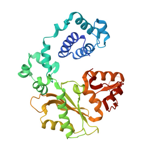
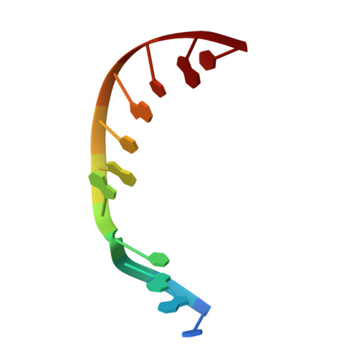
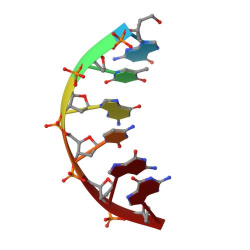
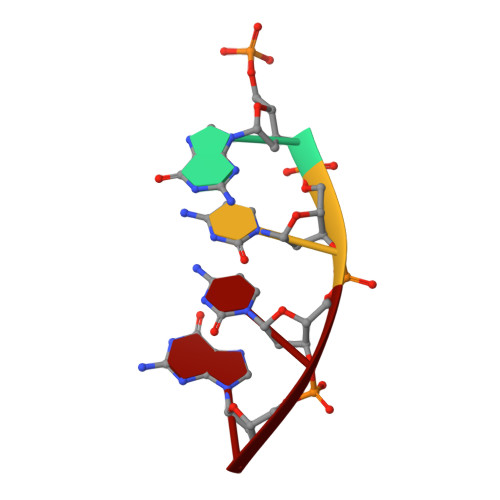
    DNA polymerase lambda (Pol lambda) is one of several DNA polymerases suggested to participate in base excision repair (BER), in repair of broken DNA ends and in translesion synthesis. It has been proposed that the nature of the DNA intermediates partly determines which polymerase is used for a particular repair reaction. To test this hypothesis, here we examine the ability of human Pol lambda to extend mismatched primer-termini, either on 'open' template-primer substrates, or on its preferred substrate, a 1 nt gapped-DNA molecule having a 5'-phosphate. Interestingly, Pol lambda extended mismatches with an average efficiency of approximately 10(-2) relative to matched base pairs. The match and mismatch extension catalytic efficiencies obtained on gapped molecules were approximately 260-fold higher than on template-primer molecules. A crystal structure of Pol lambda in complex with a single-nucleotide gap containing a dG.dGMP mismatch at the primer-terminus (2.40 A) suggests that, at least for certain mispairs, Pol lambda is unable to differentiate between matched and mismatched termini during the DNA binding step, thus accounting for the relatively high efficiency of mismatch extension. This property of Pol lambda suggests a potential role as a 'mismatch extender' during non-homologous end joining (NHEJ), and possibly during translesion synthesis.


  • Organizational Affiliation
    • Centro de Biología Molecular Severo Ochoa (CSIC-UAM), Universidad Autónoma, 28049 Madrid, Spain.

Macromolecule Content 

  • Total Structure Weight: 176.3 kDa 
  • Atom Count: 12,600 
  • Modeled Residue Count: 1,389 
  • Deposited Residue Count: 1,424 
  • Unique protein chains: 1
  • Unique nucleic acid chains: 3

Macromolecules


Find similar proteins by:|  3D Structure
Entity ID: 4
MoleculeChains  Sequence LengthOrganismDetailsImage
DNA polymerase lambdaM [auth A],
N [auth E],
O [auth I],
P [auth M]
335Homo sapiensMutation(s): 0 
Gene Names: POLL
EC: 2.7.7.7 (PDB Primary Data), 4.2.99 (PDB Primary Data)
UniProt & NIH Common Fund Data Resources
Find proteins for Q9UGP5 (Homo sapiens)
Explore Q9UGP5 
Go to UniProtKB:  Q9UGP5
PHAROS:  Q9UGP5
GTEx:  ENSG00000166169 
Entity Groups
Sequence Clusters30% Identity50% Identity70% Identity90% Identity95% Identity100% Identity
UniProt GroupQ9UGP5
Sequence Annotations
Expand
Reference Sequence
Find similar nucleic acids by:  Sequence
Entity ID: 1
MoleculeChains LengthOrganismImage
5'-D(*CP*GP*GP*CP*AP*GP*CP*GP*CP*AP*C)-3'A [auth T],
D [auth F],
G [auth J],
J [auth N]
11N/A
Sequence Annotations
Expand
Reference Sequence
Find similar nucleic acids by:  Sequence
Entity ID: 2
MoleculeChains LengthOrganismImage
5'-D(*GP*TP*GP*CP*GP*G)-3'B [auth P],
E [auth G],
H [auth K],
K [auth R]
6N/A
Sequence Annotations
Expand
Reference Sequence
Find similar nucleic acids by:  Sequence
Entity ID: 3
MoleculeChains LengthOrganismImage
5'-D(P*GP*CP*CP*G)-3'C [auth D],
F [auth H],
I [auth L],
L [auth Q]
4N/A
Sequence Annotations
Expand
Reference Sequence

Small Molecules

Ligands 5 Unique
IDChains Name / Formula / InChI Key2D Diagram3D Interactions
CAC

Query on CAC


Download:Ideal Coordinates CCD File
S [auth D],
U [auth H],
V [auth J]
CACODYLATE ION
C2 H6 As O2
OGGXGZAMXPVRFZ-UHFFFAOYSA-M
EDO

Query on EDO


Download:Ideal Coordinates CCD File
BA [auth E],
W [auth N]
1,2-ETHANEDIOL
C2 H6 O2
LYCAIKOWRPUZTN-UHFFFAOYSA-N
CL

Query on CL


Download:Ideal Coordinates CCD File
Z [auth A]CHLORIDE ION
Cl
VEXZGXHMUGYJMC-UHFFFAOYSA-M
MG

Query on MG


Download:Ideal Coordinates CCD File
T [auth F]MAGNESIUM ION
Mg
JLVVSXFLKOJNIY-UHFFFAOYSA-N
NA

Query on NA


Download:Ideal Coordinates CCD File
AA [auth E]
CA [auth I]
DA [auth M]
EA [auth M]
Q [auth T]
AA [auth E],
CA [auth I],
DA [auth M],
EA [auth M],
Q [auth T],
R [auth P],
X [auth Q],
Y [auth A]
SODIUM ION
Na
FKNQFGJONOIPTF-UHFFFAOYSA-N

Experimental Data & Validation

Experimental Data

  • Method: X-RAY DIFFRACTION
  • Resolution: 2.40 Å
  • R-Value Free:  0.253 (Depositor) 
  • R-Value Work:  0.209 (Depositor), 0.201 (DCC) 
Space Group: P 21 21 2
Unit Cell:
Length ( Å )Angle ( ˚ )
a = 192.374α = 90
b = 98.268β = 90
c = 104.96γ = 90
Software Package:
Software NamePurpose
DENZOdata reduction
SCALEPACKdata scaling
CNSrefinement
PDB_EXTRACTdata extraction

Structure Validation

View Full Validation Report


Entry History 

Deposition Data

Revision History  (Full details and data files)

  • Version 1.0: 2006-09-05
    Type: Initial release
  • Version 1.1: 2008-05-01
    Changes: Version format compliance
  • Version 1.2: 2011-07-13
    Changes: Non-polymer description, Version format compliance
  • Version 1.3: 2017-10-18
    Changes: Refinement description
  • Version 1.4: 2023-08-30
    Changes: Data collection, Database references, Derived calculations, Refinement description
  • Version 1.5: 2024-11-13
    Changes: Structure summary