2H7R | pdb_00002h7r

L244A mutant of Cytochrome P450cam complexed with imidazole


Experimental Data Snapshot

  • Method: X-RAY DIFFRACTION
  • Resolution: 2.10 Å
  • R-Value Free: 
    0.283 (Depositor), 0.280 (DCC) 
  • R-Value Work: 
    0.210 (Depositor), 0.213 (DCC) 
  • R-Value Observed: 
    0.210 (Depositor) 

Starting Model: experimental
View more details

wwPDB Validation 3D Report Full Report

Validation slider image for 2H7R

Ligand Structure Quality Assessment 


This is version 1.4 of the entry. See complete history

Literature

Cytochrome P450 active site plasticity: attenuation of imidazole binding in cytochrome P450cam by an L244A mutation.

Verras, A.Alian, A.Montellano, P.R.

(2006) Protein Eng Des Sel 19: 491-496

  • DOI: https://doi.org/10.1093/protein/gzl035
  • Primary Citation Related Structures: 
    2H7Q, 2H7R, 2H7S

  • PubMed Abstract: 

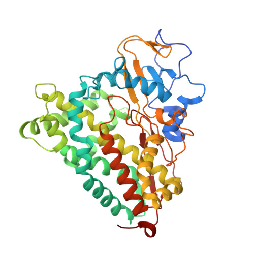
    We have identified a P450(cam) mutation, L244A, that mitigates the affinity for imidazole and substituted imidazoles while maintaining a high affinity for the natural substrate camphor. The P450(cam) L244A crystal structure solved in the absence of any ligand reveals that the I-helix is displaced inwards by over 1 A in response to the cavity created by the change from leucine to alanine. Furthermore, the crystal structures of imidazole-bound P450(cam) and the 1-methylimidazole-bound P450(cam) L244A mutant reveal that the ligands have distinct binding modes in the two proteins. Whereas in wild-type P450(cam) the imidazole coordinates to the iron in an orientation roughly perpendicular to the plane of the heme, in the L244A mutant the rearranged I helix, and specifically residue Val247, forces the imidazole into an orientation almost parallel to the heme that impairs its ability to coordinate to the heme iron. As a result, the imidazole is much more weakly bound to the mutant than it is to the wild-type enzyme. Despite the constriction of the active site by the mutation, previous work with the L244A mutant has shown that it oxidizes larger substrates than the wild-type enzyme. This paradoxical situation, in which a mutation that nominally increases the active site cavity appears to decrease it, suggests that the mutation actually increases the active site maleability, allowing it to better expand to oxidize larger substrates.


  • Organizational Affiliation
    • Department of Pharmaceutical Chemistry, University of California 600 16th Street, San Francisco, CA 94158-2517, USA.

Macromolecule Content 

  • Total Structure Weight: 47.21 kDa 
  • Atom Count: 3,359 
  • Modeled Residue Count: 405 
  • Deposited Residue Count: 414 
  • Unique protein chains: 1

Macromolecules

Find similar proteins by:|  3D Structure
Entity ID: 1
MoleculeChains  Sequence LengthOrganismDetailsImage
Cytochrome P450-cam414Pseudomonas putidaMutation(s): 2 
Gene Names: camCcyp101
EC: 1.14.15.1
UniProt
Find proteins for P00183 (Pseudomonas putida)
Explore P00183 
Go to UniProtKB:  P00183
Entity Groups
Sequence Clusters30% Identity50% Identity70% Identity90% Identity95% Identity100% Identity
UniProt GroupP00183
Sequence Annotations
Expand
Reference Sequence

Small Molecules

Ligands 2 Unique
IDChains Name / Formula / InChI Key2D Diagram3D Interactions
HEM

Query on HEM


Download:Ideal Coordinates CCD File
B [auth A]PROTOPORPHYRIN IX CONTAINING FE
C34 H32 Fe N4 O4
KABFMIBPWCXCRK-RGGAHWMASA-L
1MZ

Query on 1MZ


Download:Ideal Coordinates CCD File
C [auth A]1-METHYLIMIDAZOLE
C4 H7 N2
MCTWTZJPVLRJOU-UHFFFAOYSA-O

Experimental Data & Validation

Experimental Data

  • Method: X-RAY DIFFRACTION
  • Resolution: 2.10 Å
  • R-Value Free:  0.283 (Depositor), 0.280 (DCC) 
  • R-Value Work:  0.210 (Depositor), 0.213 (DCC) 
  • R-Value Observed: 0.210 (Depositor) 
Space Group: P 1 21 1
Unit Cell:
Length ( Å )Angle ( ˚ )
a = 56.599α = 90
b = 58.784β = 105.29
c = 57.441γ = 90
Software Package:
Software NamePurpose
DENZOdata reduction
CNSrefinement
SCALEPACKdata scaling
CNSphasing

Structure Validation

View Full Validation Report


Ligand Structure Quality Assessment 


Entry History 

Deposition Data

Revision History  (Full details and data files)

  • Version 1.0: 2006-10-31
    Type: Initial release
  • Version 1.1: 2007-10-16
    Changes: Version format compliance
  • Version 1.2: 2011-07-13
    Changes: Version format compliance
  • Version 1.3: 2021-10-20
    Changes: Database references, Derived calculations
  • Version 1.4: 2023-08-30
    Changes: Data collection, Refinement description