2HC4 | pdb_00002hc4

Crystal structure of the LBD of VDR of Danio rerio in complex with calcitriol


Experimental Data Snapshot

  • Method: X-RAY DIFFRACTION
  • Resolution: 2.20 Å
  • R-Value Free: 
    0.250 (Depositor), 0.250 (DCC) 
  • R-Value Work: 
    0.220 (Depositor), 0.220 (DCC) 
  • R-Value Observed: 
    0.220 (Depositor) 

Starting Model: experimental
View more details

wwPDB Validation   3D Report Full Report


Ligand Structure Quality Assessment 

Created with Raphaël 2.3.0Worse 01 BetterLigand structure goodness of fit to experimental dataBest fitted VDXClick on this verticalbar to view details

This is version 1.4 of the entry. See complete history


Literature

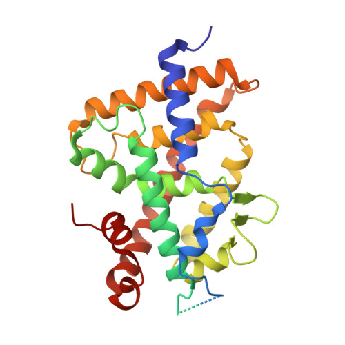
Adaptability of the Vitamin D nuclear receptor to the synthetic ligand Gemini: remodelling the LBP with one side chain rotation

Ciesielski, F.Rochel, N.Moras, D.

(2007) J Steroid Biochem Mol Biol 103: 235-242

  • DOI: https://doi.org/10.1016/j.jsbmb.2006.12.003
  • Primary Citation of Related Structures:  
    2HC4, 2HCD

  • PubMed Abstract: 

    The crystal structure of the ligand binding domain (LBD) of the wild-type Vitamin D receptor (VDR) of zebrafish bound to Gemini, a synthetic agonist ligand with two identical side chains branching at carbon 20 reveals a ligand-dependent structural rearrangement of the ligand binding pocket (LBP). The rotation of a Leu side chain opens the access to a channel that can accommodate the second side chain of the ligand. The 25% increase of the LBP's volume does not alter the essential agonist features of VDR. The possibility to adapt the LBP to novel ligands with different chemistry and/or structure opens new perspectives in the design of more specifically targeted ligands.


  • Organizational Affiliation

    Département de Biologie et de Génomique Structurales, IGBMC, CNRS/INSERM Université Louis Pasteur, Parc d'innovation BP10142, 67404 Illkirch Cedex, France.


Macromolecules
Find similar proteins by:  (by identity cutoff)  |  3D Structure
Entity ID: 1
MoleculeChains Sequence LengthOrganismDetailsImage
Vitamin D receptor302Danio rerioMutation(s): 0 
UniProt
Find proteins for Q9PTN2 (Danio rerio)
Explore Q9PTN2 
Go to UniProtKB:  Q9PTN2
Entity Groups  
Sequence Clusters30% Identity50% Identity70% Identity90% Identity95% Identity100% Identity
UniProt GroupQ9PTN2
Sequence Annotations
Expand
  • Reference Sequence

Find similar proteins by:  Sequence   |   3D Structure  

Entity ID: 2
MoleculeChains Sequence LengthOrganismDetailsImage
SRC-1 from Nuclear receptor coactivator 115Homo sapiensMutation(s): 0 
EC: 2.3.1.48
UniProt & NIH Common Fund Data Resources
Find proteins for Q15788 (Homo sapiens)
Explore Q15788 
Go to UniProtKB:  Q15788
PHAROS:  Q15788
GTEx:  ENSG00000084676 
Entity Groups  
Sequence Clusters30% Identity50% Identity70% Identity90% Identity95% Identity100% Identity
UniProt GroupQ15788
Sequence Annotations
Expand
  • Reference Sequence
Small Molecules
Ligands 1 Unique
IDChains Name / Formula / InChI Key2D Diagram3D Interactions
VDX
Query on VDX

Download Ideal Coordinates CCD File 
C [auth A]5-{2-[1-(5-HYDROXY-1,5-DIMETHYL-HEXYL)-7A-METHYL-OCTAHYDRO-INDEN-4-YLIDENE]-ETHYLIDENE}-4-METHYLENE-CYCLOHEXANE-1,3-DIOL
C27 H44 O3
GMRQFYUYWCNGIN-NKMMMXOESA-N
Binding Affinity Annotations 
IDSourceBinding Affinity
VDX BindingDB:  2HC4 Kd: 1200 (nM) from 1 assay(s)
EC50: 5.5 (nM) from 1 assay(s)
Experimental Data & Validation

Experimental Data

  • Method: X-RAY DIFFRACTION
  • Resolution: 2.20 Å
  • R-Value Free:  0.250 (Depositor), 0.250 (DCC) 
  • R-Value Work:  0.220 (Depositor), 0.220 (DCC) 
  • R-Value Observed: 0.220 (Depositor) 
Space Group: P 65 2 2
Unit Cell:
Length ( Å )Angle ( ˚ )
a = 65.835α = 90
b = 65.835β = 90
c = 264.839γ = 120
Software Package:
Software NamePurpose
CNSrefinement
ADSCdata collection
HKL-2000data scaling
CNSphasing

Structure Validation

View Full Validation Report


Ligand Structure Quality Assessment 

Created with Raphaël 2.3.0Worse 01 BetterLigand structure goodness of fit to experimental dataBest fitted VDXClick on this verticalbar to view details

Entry History 

Deposition Data

Revision History  (Full details and data files)

  • Version 1.0: 2007-05-01
    Type: Initial release
  • Version 1.1: 2008-05-01
    Changes: Version format compliance
  • Version 1.2: 2011-07-13
    Changes: Version format compliance
  • Version 1.3: 2021-03-03
    Changes: Advisory, Database references, Derived calculations, Source and taxonomy
  • Version 1.4: 2023-10-25
    Changes: Advisory, Data collection, Database references, Refinement description