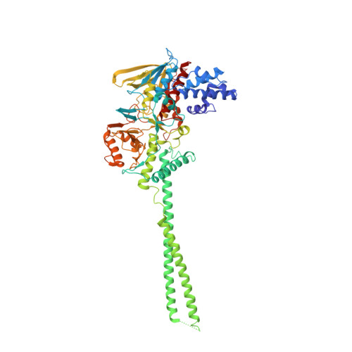
2HKO

Crystal structure of LSD1


Experimental Data Snapshot

  • Method: X-RAY DIFFRACTION
  • Resolution: 2.80 Å
  • R-Value Free: 0.283 
  • R-Value Work: 0.230 
  • R-Value Observed: 0.230 

wwPDB Validation   3D Report Full Report


Ligand Structure Quality Assessment 


This is version 1.4 of the entry. See complete history


Literature

Crystal structure of human histone lysine-specific demethylase 1 (LSD1).

Chen, Y.Yang, Y.Wang, F.Wan, K.Yamane, K.Zhang, Y.Lei, M.

(2006) Proc Natl Acad Sci U S A 103: 13956-13961

  • DOI: https://doi.org/10.1073/pnas.0606381103
  • Primary Citation of Related Structures:  
    2HKO

  • PubMed Abstract: 

    Lysine-specific demethylase 1 (LSD1) was recently identified as the first histone demethylase that specifically demethylates monomethylated and dimethylated histone H3 at K4. It is a component of the CoREST and other corepressor complexes and plays an important role in silencing neuronal-specific genes in nonneuronal cells, but the molecular mechanisms of its action remain unclear. The 2.8-A-resolution crystal structure of the human LSD1 reveals that LSD1 defines a new subfamily of FAD-dependent oxidases. The active center of LSD1 is characterized by a remarkable 1,245-A3 substrate-binding cavity with a highly negative electrostatic potential. Although the protein core of LSD1 resembles other flavoenzymes, its enzymatic activity and functions require two additional structural modules: an N-terminal SWIRM domain important for protein stability and a large insertion in the catalytic domain indispensable both for the demethylase activity and the interaction with CoREST. These results provide a framework for further probing the catalytic mechanism and the functional roles of LSD1.


  • Organizational Affiliation

    Department of Biological Chemistry, University of Michigan Medical School, 5413 Medical Science I, 1301 Catherine Road, Ann Arbor, MI 48109-0606, USA.


Macromolecules
Find similar proteins by:  (by identity cutoff)  |  3D Structure
Entity ID: 1
MoleculeChains Sequence LengthOrganismDetailsImage
Lysine-specific histone demethylase 1664Homo sapiensMutation(s): 1 
Gene Names: AOF2
EC: 1 (PDB Primary Data), 1.14.99.66 (UniProt)
UniProt & NIH Common Fund Data Resources
Find proteins for O60341 (Homo sapiens)
Explore O60341 
Go to UniProtKB:  O60341
PHAROS:  O60341
GTEx:  ENSG00000004487 
Entity Groups  
Sequence Clusters30% Identity50% Identity70% Identity90% Identity95% Identity100% Identity
UniProt GroupO60341
Sequence Annotations
Expand
  • Reference Sequence
Small Molecules
Ligands 1 Unique
IDChains Name / Formula / InChI Key2D Diagram3D Interactions
FAD
Query on FAD

Download Ideal Coordinates CCD File 
B [auth A]FLAVIN-ADENINE DINUCLEOTIDE
C27 H33 N9 O15 P2
VWWQXMAJTJZDQX-UYBVJOGSSA-N
Experimental Data & Validation

Experimental Data

  • Method: X-RAY DIFFRACTION
  • Resolution: 2.80 Å
  • R-Value Free: 0.283 
  • R-Value Work: 0.230 
  • R-Value Observed: 0.230 
  • Space Group: P 61 2 2
Unit Cell:
Length ( Å )Angle ( ˚ )
a = 187.113α = 90
b = 187.113β = 90
c = 106.581γ = 120
Software Package:
Software NamePurpose
SHARPphasing
CNSrefinement
HKL-2000data reduction
HKL-2000data scaling

Structure Validation

View Full Validation Report


Ligand Structure Quality Assessment 


Entry History 

Deposition Data

Revision History  (Full details and data files)

  • Version 1.0: 2006-08-29
    Type: Initial release
  • Version 1.1: 2008-05-01
    Changes: Version format compliance
  • Version 1.2: 2011-07-13
    Changes: Version format compliance
  • Version 1.3: 2021-10-20
    Changes: Database references, Derived calculations
  • Version 1.4: 2024-02-14
    Changes: Data collection