2HQW

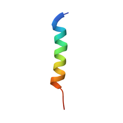
Crystal Structure of Ca2+/Calmodulin bound to NMDA Receptor NR1C1 peptide


Experimental Data Snapshot

  • Method: X-RAY DIFFRACTION
  • Resolution: 1.90 Å
  • R-Value Free: 0.248 
  • R-Value Work: 0.206 
  • R-Value Observed: 0.207 

wwPDB Validation   3D Report Full Report


This is version 1.2 of the entry. See complete history


Literature

The NMDA Receptor NR1 C1 Region Bound to Calmodulin: Structural Insights into Functional Differences between Homologous Domains.

Ataman, Z.A.Gakhar, L.Sorensen, B.R.Hell, J.W.Shea, M.A.

(2007) Structure 15: 1603-1617

  • DOI: https://doi.org/10.1016/j.str.2007.10.012
  • Primary Citation of Related Structures:  
    2HQW

  • PubMed Abstract: 

    Calmodulin (CaM) regulates tetrameric N-methyl-D-aspartate receptors (NMDARs) by binding tightly to the C0 and C1 regions of its NR1 subunit. A crystal structure (2HQW; 1.96 A) of calcium-saturated CaM bound to NR1C1 (peptide spanning 875-898) showed that NR1 S890, whose phosphorylation regulates membrane localization, was solvent protected, whereas the endoplasmic reticulum retention motif was solvent exposed. NR1 F880 filled the CaM C-domain pocket, whereas T886 was closest to the N-domain pocket. This 1-7 pattern was most similar to that in the CaM-MARCKS complex. Comparison of CaM-ligand wrap-around conformations identified a core tetrad of CaM C-domain residues (FLMM(C)) that contacted all ligands consistently. An identical tetrad of N-domain residues (FLMM(N)) made variable sets of contacts with ligands. This CaM-NR1C1 structure provides a foundation for designing mutants to test the role of CaM in NR1 trafficking as well as insights into how the homologous CaM domains have different roles in molecular recognition.


  • Organizational Affiliation

    Department of Biochemistry, Roy J. and Lucille A. Carver College of Medicine, University of Iowa, Iowa City, IA 52242-1109, USA.


Macromolecules
Find similar proteins by:  (by identity cutoff)  |  3D Structure
Entity ID: 1
MoleculeChains Sequence LengthOrganismDetailsImage
Calmodulin148Rattus norvegicusMutation(s): 0 
Gene Names: Calm1CalmCamCam1
UniProt
Find proteins for P0DP29 (Rattus norvegicus)
Explore P0DP29 
Go to UniProtKB:  P0DP29
Entity Groups  
Sequence Clusters30% Identity50% Identity70% Identity90% Identity95% Identity100% Identity
UniProt GroupP0DP29
Sequence Annotations
Expand
  • Reference Sequence

Find similar proteins by:  Sequence   |   3D Structure  

Entity ID: 2
MoleculeChains Sequence LengthOrganismDetailsImage
Glutamate NMDA receptor subunit zeta 124N/AMutation(s): 0 
UniProt
Find proteins for P35439 (Rattus norvegicus)
Explore P35439 
Go to UniProtKB:  P35439
Entity Groups  
Sequence Clusters30% Identity50% Identity70% Identity90% Identity95% Identity100% Identity
UniProt GroupP35439
Sequence Annotations
Expand
  • Reference Sequence
Experimental Data & Validation

Experimental Data

  • Method: X-RAY DIFFRACTION
  • Resolution: 1.90 Å
  • R-Value Free: 0.248 
  • R-Value Work: 0.206 
  • R-Value Observed: 0.207 
  • Space Group: P 32 2 1
Unit Cell:
Length ( Å )Angle ( ˚ )
a = 40.371α = 90
b = 40.371β = 90
c = 175.765γ = 120
Software Package:
Software NamePurpose
REFMACrefinement
AMoREphasing

Structure Validation

View Full Validation Report


Entry History 

Deposition Data

Revision History  (Full details and data files)

  • Version 1.0: 2007-11-13
    Type: Initial release
  • Version 1.1: 2011-07-13
    Changes: Derived calculations, Version format compliance
  • Version 1.2: 2024-02-14
    Changes: Data collection, Database references, Derived calculations