2HV7 | pdb_00002hv7

Crystal structure of phosphotyrosyl phosphatase activator bound to ATPgammaS


Experimental Data Snapshot

  • Method: X-RAY DIFFRACTION
  • Resolution: 2.50 Å
  • R-Value Free: 
    0.312 (Depositor) 
  • R-Value Work: 
    0.249 (Depositor) 
  • R-Value Observed: 
    0.252 (Depositor) 

Starting Model: experimental
View more details

wwPDB Validation 3D Report Full Report

Validation slider image for 2HV7

This is version 1.3 of the entry. See complete history

Literature

Structure and mechanism of the phosphotyrosyl phosphatase activator.

Chao, Y.Xing, Y.Chen, Y.Xu, Y.Lin, Z.Li, Z.Jeffrey, P.D.Stock, J.B.Shi, Y.

(2006) Mol Cell 23: 535-546

  • DOI: https://doi.org/10.1016/j.molcel.2006.07.027
  • Primary Citation Related Structures: 
    2HV6, 2HV7

  • PubMed Abstract: 

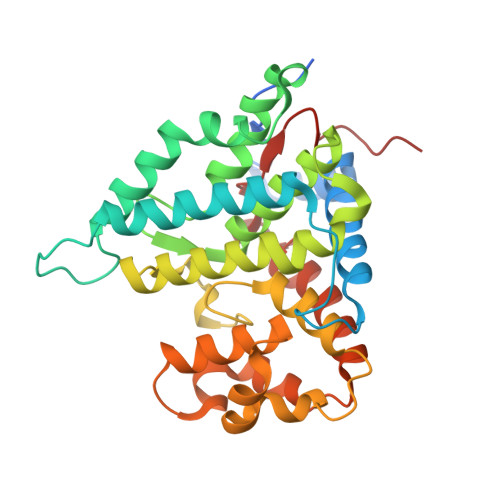
    Phosphotyrosyl phosphatase activator (PTPA), also known as PP2A phosphatase activator, is a conserved protein from yeast to human. Here we report the 1.9 A crystal structure of human PTPA, which reveals a previously unreported fold consisting of three subdomains: core, lid, and linker. Structural analysis uncovers a highly conserved surface patch, which borders the three subdomains, and an associated deep pocket located between the core and the linker subdomains. The conserved surface patch and the deep pocket are responsible for binding to PP2A and ATP, respectively. PTPA and PP2A A-C dimer together constitute a composite ATPase. PTPA binding to PP2A results in a dramatic alteration of substrate specificity, with enhanced phosphotyrosine phosphatase activity and decreased phosphoserine phosphatase activity. This function of PTPA strictly depends on the composite ATPase activity. These observations reveal significant insights into the function and mechanism of PTPA and have important ramifications for understanding PP2A function.


  • Organizational Affiliation
    • Department of Molecular Biology, Lewis Thomas Laboratory, Princeton University, Princeton, New Jersey 08544, USA.

Macromolecule Content 

  • Total Structure Weight: 296.26 kDa 
  • Atom Count: 19,612 
  • Modeled Residue Count: 2,408 
  • Deposited Residue Count: 2,584 
  • Unique protein chains: 1

Macromolecules

Find similar proteins by:|  3D Structure
Entity ID: 1
MoleculeChains  Sequence LengthOrganismDetailsImage
Protein phosphatase 2A, regulatory subunit B
A, B, C, D, E
A, B, C, D, E, F, G, H
323Homo sapiensMutation(s): 0 
Gene Names: PPP2R4
EC: 5.2.1.8
UniProt & NIH Common Fund Data Resources
Find proteins for Q15257 (Homo sapiens)
Explore Q15257 
Go to UniProtKB:  Q15257
PHAROS:  Q15257
GTEx:  ENSG00000119383 
Entity Groups
Sequence Clusters30% Identity50% Identity70% Identity90% Identity95% Identity100% Identity
UniProt GroupQ15257
Sequence Annotations
Expand
Reference Sequence

Experimental Data & Validation

Experimental Data

  • Method: X-RAY DIFFRACTION
  • Resolution: 2.50 Å
  • R-Value Free:  0.312 (Depositor) 
  • R-Value Work:  0.249 (Depositor) 
  • R-Value Observed: 0.252 (Depositor) 
Space Group: P 1
Unit Cell:
Length ( Å )Angle ( ˚ )
a = 80.041α = 73.91
b = 86.096β = 89.97
c = 95.099γ = 73.31
Software Package:
Software NamePurpose
REFMACrefinement
HKL-2000data reduction
HKL-2000data scaling
PHASERphasing

Structure Validation

View Full Validation Report


Entry History 

Deposition Data

Revision History  (Full details and data files)

  • Version 1.0: 2006-08-22
    Type: Initial release
  • Version 1.1: 2008-05-01
    Changes: Version format compliance
  • Version 1.2: 2011-07-13
    Changes: Version format compliance
  • Version 1.3: 2023-08-30
    Changes: Data collection, Database references, Derived calculations, Refinement description