2HZI

Abl kinase domain in complex with PD180970


Experimental Data Snapshot

  • Method: X-RAY DIFFRACTION
  • Resolution: 1.70 Å
  • R-Value Free: 0.204 
  • R-Value Work: 0.175 
  • R-Value Observed: 0.177 

wwPDB Validation   3D Report Full Report


Ligand Structure Quality Assessment 


This is version 1.3 of the entry. See complete history


Literature

Structural biology contributions to the discovery of drugs to treat chronic myelogenous leukaemia.

Cowan-Jacob, S.W.Fendrich, G.Floersheimer, A.Furet, P.Liebetanz, J.Rummel, G.Rheinberger, P.Centeleghe, M.Fabbro, D.Manley, P.W.

(2007) Acta Crystallogr D Biol Crystallogr 63: 80-93

  • DOI: https://doi.org/10.1107/S0907444906047287
  • Primary Citation of Related Structures:  
    2HYY, 2HZ0, 2HZ4, 2HZI, 2HZN

  • PubMed Abstract: 

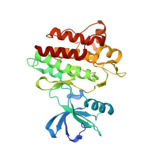
    Chronic myelogenous leukaemia (CML) results from the Bcr-Abl oncoprotein, which has a constitutively activated Abl tyrosine kinase domain. Although most chronic phase CML patients treated with imatinib as first-line therapy maintain excellent durable responses, patients who have progressed to advanced-stage CML frequently fail to respond or lose their response to therapy owing to the emergence of drug-resistant mutants of the protein. More than 40 such point mutations have been observed in imatinib-resistant patients. The crystal structures of wild-type and mutant Abl kinase in complex with imatinib and other small-molecule Abl inhibitors were determined, with the aim of understanding the molecular basis of resistance and to aid in the design and optimization of inhibitors active against the resistance mutants. These results are presented in a way which illustrates the approaches used to generate multiple structures, the type of information that can be gained and the way that this information is used to support drug discovery.


  • Organizational Affiliation

    Novartis Institutes for Biomedical Research, Basel, Switzerland. sandra.jacob@novartis.com


Macromolecules
Find similar proteins by:  (by identity cutoff)  |  3D Structure
Entity ID: 1
MoleculeChains Sequence LengthOrganismDetailsImage
Proto-oncogene tyrosine-protein kinase ABL1
A, B
277Homo sapiensMutation(s): 0 
Gene Names: ABL1
EC: 2.7.10.2
UniProt & NIH Common Fund Data Resources
Find proteins for P00519 (Homo sapiens)
Explore P00519 
Go to UniProtKB:  P00519
PHAROS:  P00519
GTEx:  ENSG00000097007 
Entity Groups  
Sequence Clusters30% Identity50% Identity70% Identity90% Identity95% Identity100% Identity
UniProt GroupP00519
Sequence Annotations
Expand
  • Reference Sequence
Small Molecules
Ligands 1 Unique
IDChains Name / Formula / InChI Key2D Diagram3D Interactions
JIN
Query on JIN

Download Ideal Coordinates CCD File 
C [auth A],
D [auth B]
6-(2,6-DICHLOROPHENYL)-2-[(4-FLUORO-3-METHYLPHENYL)AMINO]-8-METHYLPYRIDO[2,3-D]PYRIMIDIN-7(8H)-ONE
C21 H15 Cl2 F N4 O
SLCFEJAMCRLYRG-UHFFFAOYSA-N
Binding Affinity Annotations 
IDSourceBinding Affinity
JIN BindingDB:  2HZI Kd: min: 0.7, max: 600 (nM) from 7 assay(s)
Binding MOAD:  2HZI IC50: 70 (nM) from 1 assay(s)
PDBBind:  2HZI IC50: 70 (nM) from 1 assay(s)
Experimental Data & Validation

Experimental Data

  • Method: X-RAY DIFFRACTION
  • Resolution: 1.70 Å
  • R-Value Free: 0.204 
  • R-Value Work: 0.175 
  • R-Value Observed: 0.177 
  • Space Group: P 21 21 2
Unit Cell:
Length ( Å )Angle ( ˚ )
a = 106.475α = 90
b = 131.533β = 90
c = 56.459γ = 90
Software Package:
Software NamePurpose
SCALAdata scaling
REFMACrefinement
PDB_EXTRACTdata extraction
MOSFLMdata reduction
CCP4data scaling

Structure Validation

View Full Validation Report


Ligand Structure Quality Assessment 


Entry History 

Deposition Data

Revision History  (Full details and data files)

  • Version 1.0: 2007-01-16
    Type: Initial release
  • Version 1.1: 2008-05-01
    Changes: Version format compliance
  • Version 1.2: 2011-07-13
    Changes: Version format compliance
  • Version 1.3: 2024-02-21
    Changes: Data collection, Database references, Derived calculations