2IT4 | pdb_00002it4

X ray structure of the complex between Carbonic Anhydrase I and the phosphonate antiviral drug foscarnet


Experimental Data Snapshot

  • Method: X-RAY DIFFRACTION
  • Resolution: 2.00 Å
  • R-Value Free: 
    0.316 (Depositor), 0.201 (DCC) 
  • R-Value Work: 
    0.246 (Depositor) 
  • R-Value Observed: 
    0.249 (Depositor) 

Starting Model: experimental
View more details

wwPDB Validation 3D Report Full Report

Validation slider image for 2IT4

This is version 1.5 of the entry. See complete history

Literature

Phosph(on)ate as a zinc-binding group in metalloenzyme inhibitors: X-ray crystal structure of the antiviral drug foscarnet complexed to human carbonic anhydrase I.

Temperini, C.Innocenti, A.Guerri, A.Scozzafava, A.Rusconi, S.Supuran, C.T.

(2007) Bioorg Med Chem Lett 17: 2210-2215

  • DOI: https://doi.org/10.1016/j.bmcl.2007.01.113
  • Primary Citation Related Structures: 
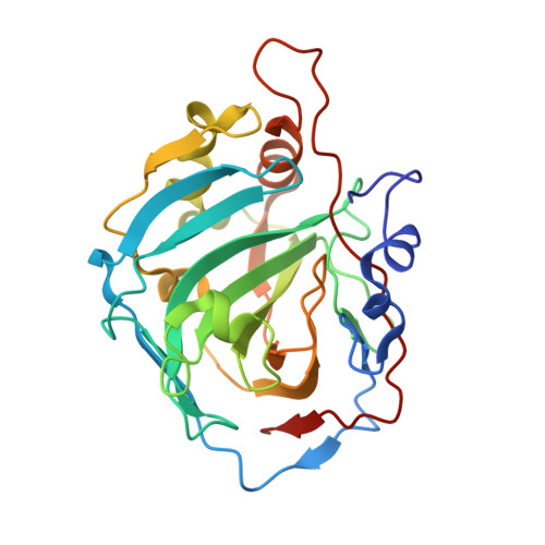
    2IT4

  • PubMed Abstract: 

    Foscarnet (phosphonoformate trisodium salt), an antiviral used for the treatment of HIV and herpes virus infections, also acts as an activator or inhibitor of the metalloenzyme carbonic anhydrase (CA, EC 4.2.1.1). Interaction of the drug with 11 CA isozymes has been investigated kinetically, and the X-ray structure of its adduct with isoform I (hCA I-foscarnet complex) has been resolved. The first CA inhibitor possessing a phosphonate zinc-binding group is thus evidenced, together with the factors governing recognition of such small molecules by a metalloenzyme active site. Foscarnet is also a clear-cut example of modulator of an enzyme activity which can act either as an activator or inhibitor of a CA isozyme.


  • Organizational Affiliation
    • Università degli Studi di Firenze, Laboratorio di Chimica Bioinorganica, Rm. 188, Via della Lastruccia 3, I-50019 Sesto Fiorentino (Florence), Italy.

Macromolecule Content 

  • Total Structure Weight: 57.07 kDa 
  • Atom Count: 4,385 
  • Modeled Residue Count: 512 
  • Deposited Residue Count: 512 
  • Unique protein chains: 1

Macromolecules

Find similar proteins by:|  3D Structure
Entity ID: 1
MoleculeChains  Sequence LengthOrganismDetailsImage
Carbonic anhydrase 1
A, B
256Homo sapiensMutation(s): 0 
EC: 4.2.1.1 (PDB Primary Data), 4.2.1.69 (UniProt)
UniProt & NIH Common Fund Data Resources
Find proteins for P00915 (Homo sapiens)
Explore P00915 
Go to UniProtKB:  P00915
PHAROS:  P00915
GTEx:  ENSG00000133742 
Entity Groups
Sequence Clusters30% Identity50% Identity70% Identity90% Identity95% Identity100% Identity
UniProt GroupP00915
Sequence Annotations
Expand
Reference Sequence

Experimental Data & Validation

Experimental Data

  • Method: X-RAY DIFFRACTION
  • Resolution: 2.00 Å
  • R-Value Free:  0.316 (Depositor), 0.201 (DCC) 
  • R-Value Work:  0.246 (Depositor) 
  • R-Value Observed: 0.249 (Depositor) 
Space Group: P 21 21 21
Unit Cell:
Length ( Å )Angle ( ˚ )
a = 62.58α = 90
b = 69.51β = 90
c = 120.44γ = 90
Software Package:
Software NamePurpose
CrysalisProdata collection
AMoREphasing
REFMACrefinement
CrysalisProdata reduction
SCALAdata scaling

Structure Validation

View Full Validation Report


Entry History 

Deposition Data

Revision History  (Full details and data files)

  • Version 1.0: 2007-09-11
    Type: Initial release
  • Version 1.1: 2011-07-13
    Changes: Version format compliance
  • Version 1.2: 2017-10-18
    Changes: Refinement description
  • Version 1.3: 2018-01-31
    Changes: Experimental preparation
  • Version 1.4: 2018-04-04
    Changes: Data collection
  • Version 1.5: 2023-08-30
    Changes: Data collection, Database references, Derived calculations, Refinement description