2IT6

Crystal Structure of DCSIGN-CRD with man2


Experimental Data Snapshot

  • Method: X-RAY DIFFRACTION
  • Resolution: 1.95 Å
  • R-Value Free: 0.241 
  • R-Value Work: 0.196 

wwPDB Validation   3D Report Full Report


This is version 2.1 of the entry. See complete history


Literature

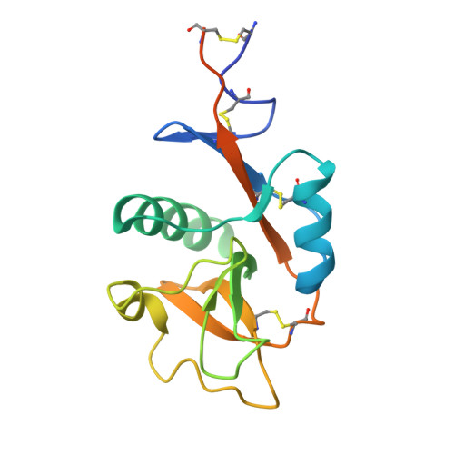
Multiple modes of binding enhance the affinity of DC-SIGN for high mannose N-linked glycans found on viral glycoproteins.

Feinberg, H.Castelli, R.Drickamer, K.Seeberger, P.H.Weis, W.I.

(2007) J Biol Chem 282: 4202-4209

  • DOI: https://doi.org/10.1074/jbc.M609689200
  • Primary Citation of Related Structures:  
    2IT5, 2IT6

  • PubMed Abstract: 

    The dendritic cell surface receptor DC-SIGN and the closely related endothelial cell receptor DC-SIGNR specifically recognize high mannose N-linked carbohydrates on viral pathogens. Previous studies have shown that these receptors bind the outer trimannose branch Manalpha1-3[Manalpha1-6]Manalpha present in high mannose structures. Although the trimannoside binds to DC-SIGN or DC-SIGNR more strongly than mannose, additional affinity enhancements are observed in the presence of one or more Manalpha1-2Manalpha moieties on the nonreducing termini of oligomannose structures. The molecular basis of this enhancement has been investigated by determining crystal structures of DC-SIGN bound to a synthetic six-mannose fragment of a high mannose N-linked oligosaccharide, Manalpha1-2Manalpha1-3[Manalpha1-2Manalpha1-6]Manalpha1-6Man and to the disaccharide Manalpha1-2Man. The structures reveal mixtures of two binding modes in each case. Each mode features typical C-type lectin binding at the principal Ca2+-binding site by one mannose residue. In addition, other sugar residues form contacts unique to each binding mode. These results suggest that the affinity enhancement displayed toward oligosaccharides decorated with the Manalpha1-2Manalpha structure is due in part to multiple binding modes at the primary Ca2+ site, which provide both additional contacts and a statistical (entropic) enhancement of binding.


  • Organizational Affiliation

    Departments of Structural Biology and Molecular & Cellular Physiology, Stanford University School of Medicine, Stanford, California 94305, USA.


Macromolecules
Find similar proteins by:  (by identity cutoff)  |  3D Structure
Entity ID: 1
MoleculeChains Sequence LengthOrganismDetailsImage
CD209 antigen155Homo sapiensMutation(s): 0 
Gene Names: CD209CLEC4L
UniProt & NIH Common Fund Data Resources
Find proteins for Q9NNX6 (Homo sapiens)
Explore Q9NNX6 
Go to UniProtKB:  Q9NNX6
PHAROS:  Q9NNX6
GTEx:  ENSG00000090659 
Entity Groups  
Sequence Clusters30% Identity50% Identity70% Identity90% Identity95% Identity100% Identity
UniProt GroupQ9NNX6
Sequence Annotations
Expand
  • Reference Sequence
Oligosaccharides

Help

Entity ID: 2
MoleculeChains Length2D Diagram Glycosylation3D Interactions
alpha-D-mannopyranose-(1-2)-alpha-D-mannopyranose
B
2N/A
Glycosylation Resources
GlyTouCan:  G53402KW
GlyCosmos:  G53402KW
GlyGen:  G53402KW
Biologically Interesting Molecules (External Reference) 1 Unique
Experimental Data & Validation

Experimental Data

  • Method: X-RAY DIFFRACTION
  • Resolution: 1.95 Å
  • R-Value Free: 0.241 
  • R-Value Work: 0.196 
  • Space Group: P 43
Unit Cell:
Length ( Å )Angle ( ˚ )
a = 55.64α = 90
b = 55.64β = 90
c = 53.2γ = 90
Software Package:
Software NamePurpose
CNSrefinement
MOSFLMdata reduction
CCP4data scaling
MOLREPphasing

Structure Validation

View Full Validation Report


Entry History 

Deposition Data

Revision History  (Full details and data files)

  • Version 1.0: 2006-12-05
    Type: Initial release
  • Version 1.1: 2008-05-01
    Changes: Version format compliance
  • Version 1.2: 2011-07-13
    Changes: Version format compliance
  • Version 2.0: 2020-07-29
    Type: Remediation
    Reason: Carbohydrate remediation
    Changes: Atomic model, Data collection, Derived calculations, Structure summary
  • Version 2.1: 2023-08-30
    Changes: Data collection, Database references, Refinement description, Structure summary