2J7A

Crystal structure of cytochrome c nitrite reductase NrfHA complex from Desulfovibrio vulgaris


Experimental Data Snapshot

  • Method: X-RAY DIFFRACTION
  • Resolution: 2.30 Å
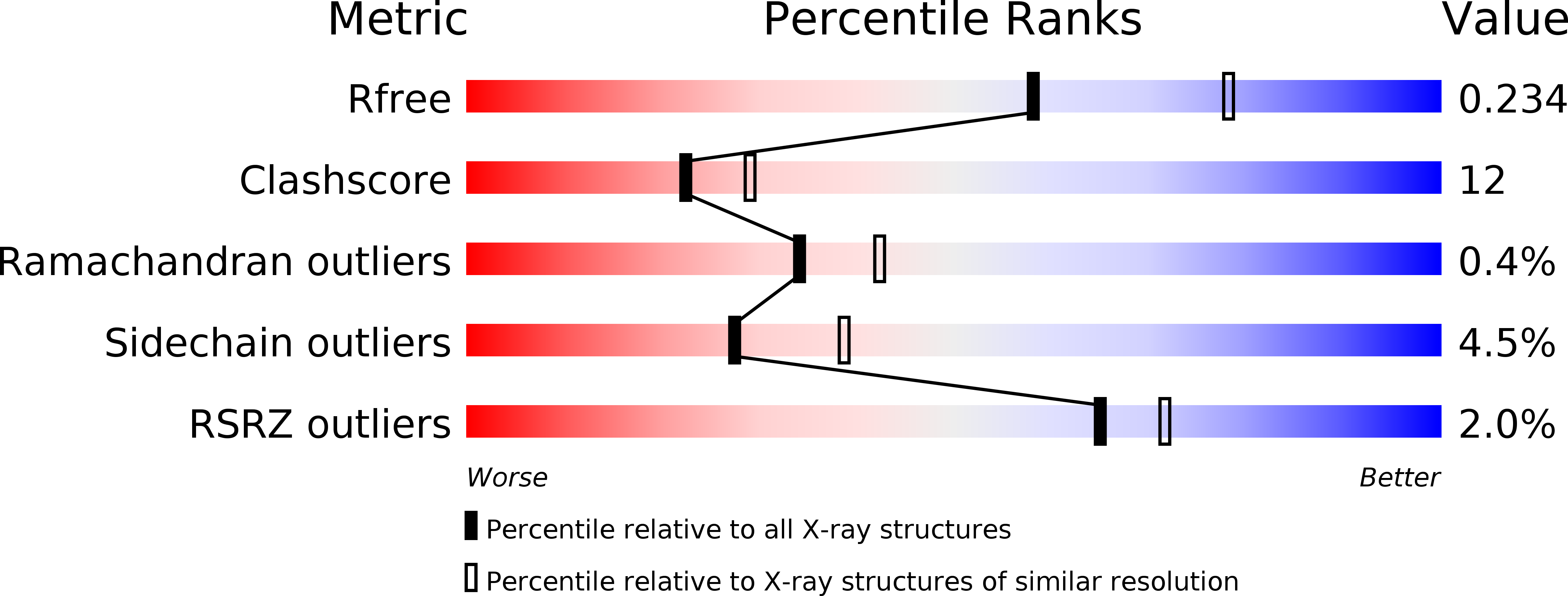
  • R-Value Free: 
    0.240 (Depositor), 0.234 (DCC) 
  • R-Value Work: 
    0.201 (Depositor), 0.197 (DCC) 
  • R-Value Observed: 
    0.203 (Depositor) 

wwPDB Validation   3D Report Full Report


Ligand Structure Quality Assessment 

Created with Raphaël 2.3.0Worse 01 BetterLigand structure goodness of fit to experimental dataBest fitted HECClick on this verticalbar to view detailsBest fitted LMTClick on this verticalbar to view details

This is version 2.2 of the entry. See complete history


Literature

X-Ray Structure of the Membrane-Bound Cytochrome C Quinol Dehydrogenase Nrfh Reveals Novel Haem Coordination.

Rodrigues, M.L.Oliveira, T.F.Pereira, I.A.C.Archer, M.

(2006) EMBO J 25: 5951

  • DOI: https://doi.org/10.1038/sj.emboj.7601439
  • Primary Citation of Related Structures:  
    2J7A

  • PubMed Abstract: 

    Oxidation of membrane-bound quinol molecules is a central step in the respiratory electron transport chains used by biological cells to generate ATP by oxidative phosphorylation. A novel family of cytochrome c quinol dehydrogenases that play an important role in bacterial respiratory chains was recognised in recent years. Here, we describe the first structure of a cytochrome from this family, NrfH from Desulfovibrio vulgaris, which forms a stable complex with its electron partner, the cytochrome c nitrite reductase NrfA. One NrfH molecule interacts with one NrfA dimer in an asymmetrical manner, forming a large membrane-bound complex with an overall alpha(4)beta(2) quaternary arrangement. The menaquinol-interacting NrfH haem is pentacoordinated, bound by a methionine from the CXXCHXM sequence, with an aspartate residue occupying the distal position. The NrfH haem that transfers electrons to NrfA has a lysine residue from the closest NrfA molecule as distal ligand. A likely menaquinol binding site, containing several conserved and essential residues, is identified.


  • Organizational Affiliation

    Instituto de Tecnologia Química e Biológica, Universidade Nova de Lisboa, ITQB-UNL, Oeiras, Portugal.


Macromolecules
Find similar proteins by:  (by identity cutoff)  |  3D Structure
Entity ID: 1
MoleculeChains Sequence LengthOrganismDetailsImage
CYTOCHROME C NITRITE REDUCTASE NRFA
A, B, D, E, G
500Nitratidesulfovibrio vulgaris str. HildenboroughMutation(s): 0 
EC: 1.7.2.2
Membrane Entity: Yes 
UniProt
Find proteins for Q72EF3 (Nitratidesulfovibrio vulgaris (strain ATCC 29579 / DSM 644 / CCUG 34227 / NCIMB 8303 / VKM B-1760 / Hildenborough))
Explore Q72EF3 
Go to UniProtKB:  Q72EF3
Entity Groups  
Sequence Clusters30% Identity50% Identity70% Identity90% Identity95% Identity100% Identity
UniProt GroupQ72EF3
Sequence Annotations
Expand
  • Reference Sequence
Find similar proteins by:  (by identity cutoff)  |  3D Structure
Entity ID: 2
MoleculeChains Sequence LengthOrganismDetailsImage
CYTOCHROME C QUINOL DEHYDROGENASE NRFH
C, F, I, L, O
159Nitratidesulfovibrio vulgaris str. HildenboroughMutation(s): 0 
Membrane Entity: Yes 
UniProt
Find proteins for Q72EF4 (Nitratidesulfovibrio vulgaris (strain ATCC 29579 / DSM 644 / CCUG 34227 / NCIMB 8303 / VKM B-1760 / Hildenborough))
Explore Q72EF4 
Go to UniProtKB:  Q72EF4
Entity Groups  
Sequence Clusters30% Identity50% Identity70% Identity90% Identity95% Identity100% Identity
UniProt GroupQ72EF4
Sequence Annotations
Expand
  • Reference Sequence
Small Molecules
Ligands 4 Unique
IDChains Name / Formula / InChI Key2D Diagram3D Interactions
HEC
Query on HEC

Download Ideal Coordinates CCD File 
AA [auth B]
AB [auth F]
AC [auth J]
AD [auth N]
AE [auth R]
HEME C
C34 H34 Fe N4 O4
HXQIYSLZKNYNMH-LJNAALQVSA-N
LMT
Query on LMT

Download Ideal Coordinates CCD File 
EB [auth F]
EE [auth R]
KA [auth C]
KD [auth O]
QC [auth L]
DODECYL-BETA-D-MALTOSIDE
C24 H46 O11
NLEBIOOXCVAHBD-QKMCSOCLSA-N
ACT
Query on ACT

Download Ideal Coordinates CCD File 
LA [auth C],
LD [auth O],
YB [auth I]
ACETATE ION
C2 H3 O2
QTBSBXVTEAMEQO-UHFFFAOYSA-M
CA
Query on CA

Download Ideal Coordinates CCD File 
EA [auth B]
EC [auth J]
ED [auth N]
FA [auth B]
FC [auth J]
CALCIUM ION
Ca
BHPQYMZQTOCNFJ-UHFFFAOYSA-N
Experimental Data & Validation

Experimental Data

  • Method: X-RAY DIFFRACTION
  • Resolution: 2.30 Å
  • R-Value Free:  0.240 (Depositor), 0.234 (DCC) 
  • R-Value Work:  0.201 (Depositor), 0.197 (DCC) 
  • R-Value Observed: 0.203 (Depositor) 
Space Group: P 21 21 21
Unit Cell:
Length ( Å )Angle ( ˚ )
a = 79.664α = 90
b = 258.117β = 90
c = 580.742γ = 90
Software Package:
Software NamePurpose
REFMACrefinement
MOSFLMdata reduction
SCALAdata scaling
SHARPphasing

Structure Validation

View Full Validation Report


Ligand Structure Quality Assessment 

Created with Raphaël 2.3.0Worse 01 BetterLigand structure goodness of fit to experimental dataBest fitted HECClick on this verticalbar to view detailsBest fitted LMTClick on this verticalbar to view details

Entry History 

Deposition Data

Revision History  (Full details and data files)

  • Version 1.0: 2006-12-06
    Type: Initial release
  • Version 2.0: 2019-09-25
    Changes: Advisory, Atomic model, Data collection, Derived calculations, Experimental preparation, Non-polymer description, Other, Structure summary
  • Version 2.1: 2019-11-06
    Changes: Data collection, Refinement description
  • Version 2.2: 2024-11-06
    Changes: Data collection, Database references, Derived calculations, Structure summary