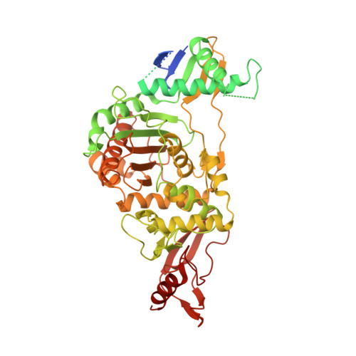
2JCH

Structural and mechanistic basis of penicillin binding protein inhibition by lactivicins


Experimental Data Snapshot

  • Method: X-RAY DIFFRACTION
  • Resolution: 2.40 Å
  • R-Value Free: 0.262 
  • R-Value Work: 0.203 
  • R-Value Observed: 0.206 

Starting Model: experimental
View more details

wwPDB Validation   3D Report Full Report


Ligand Structure Quality Assessment 


This is version 1.4 of the entry. See complete history


Literature

Structural and Mechanistic Basis of Penicillin-Binding Protein Inhibition by Lactivicins

Macheboeuf, P.Fisher, D.S.Brown, T.J.Zervosen, A.Luxen, A.Joris, B.Dessen, A.Schofield, C.J.

(2007) Nat Chem Biol 3: 565

  • DOI: https://doi.org/10.1038/nchembio.2007.21
  • Primary Citation of Related Structures:  
    2JCH, 2JE5

  • PubMed Abstract: 

    Beta-lactam antibiotics, including penicillins and cephalosporins, inhibit penicillin-binding proteins (PBPs), which are essential for bacterial cell wall biogenesis. Pathogenic bacteria have evolved efficient antibiotic resistance mechanisms that, in Gram-positive bacteria, include mutations to PBPs that enable them to avoid beta-lactam inhibition. Lactivicin (LTV; 1) contains separate cycloserine and gamma-lactone rings and is the only known natural PBP inhibitor that does not contain a beta-lactam. Here we show that LTV and a more potent analog, phenoxyacetyl-LTV (PLTV; 2), are active against clinically isolated, penicillin-resistant Streptococcus pneumoniae strains. Crystallographic analyses of S. pneumoniae PBP1b reveal that LTV and PLTV inhibition involves opening of both monocyclic cycloserine and gamma-lactone rings. In PBP1b complexes, the ring-derived atoms from LTV and PLTV show a notable structural convergence with those derived from a complexed cephalosporin (cefotaxime; 3). The structures imply that derivatives of LTV will be useful in the search for new antibiotics with activity against beta-lactam-resistant bacteria.


  • Organizational Affiliation

    Institut de Biologie Structurale Jean-Pierre Ebel Commissariat à l'énergie atomique - Centre National de La Recherche Scientifique - Université Joseph Fourier, 41 rue Jules Horowitz, F-38027 Grenoble, France.


Macromolecules
Find similar proteins by:  (by identity cutoff)  |  3D Structure
Entity ID: 1
MoleculeChains Sequence LengthOrganismDetailsImage
PENICILLIN-BINDING PROTEIN 1B720Streptococcus pneumoniae R6Mutation(s): 7 
EC: 2.4.99.28
UniProt
Find proteins for O70038 (Streptococcus pneumoniae)
Explore O70038 
Go to UniProtKB:  O70038
Entity Groups  
Sequence Clusters30% Identity50% Identity70% Identity90% Identity95% Identity100% Identity
UniProt GroupO70038
Sequence Annotations
Expand
  • Reference Sequence
Small Molecules
Ligands 4 Unique
IDChains Name / Formula / InChI Key2D Diagram3D Interactions
PL7
Query on PL7

Download Ideal Coordinates CCD File 
E [auth A](2E)-2-({(2S)-2-CARBOXY-2-[(PHENOXYACETYL)AMINO]ETHOXY}IMINO)PENTANEDIOIC ACID
C16 H18 N2 O9
LDNKNKRRFZRLIG-HWQJWEFDSA-N
SO4
Query on SO4

Download Ideal Coordinates CCD File 
B [auth A]SULFATE ION
O4 S
QAOWNCQODCNURD-UHFFFAOYSA-L
EDO
Query on EDO

Download Ideal Coordinates CCD File 
D [auth A]1,2-ETHANEDIOL
C2 H6 O2
LYCAIKOWRPUZTN-UHFFFAOYSA-N
CL
Query on CL

Download Ideal Coordinates CCD File 
C [auth A]CHLORIDE ION
Cl
VEXZGXHMUGYJMC-UHFFFAOYSA-M
Experimental Data & Validation

Experimental Data

  • Method: X-RAY DIFFRACTION
  • Resolution: 2.40 Å
  • R-Value Free: 0.262 
  • R-Value Work: 0.203 
  • R-Value Observed: 0.206 
  • Space Group: C 2 2 21
Unit Cell:
Length ( Å )Angle ( ˚ )
a = 105.84α = 90
b = 143.58β = 90
c = 97.44γ = 90
Software Package:
Software NamePurpose
REFMACrefinement
XDSdata reduction
XDSdata scaling
AMoREphasing

Structure Validation

View Full Validation Report


Ligand Structure Quality Assessment 


Entry History 

Deposition Data

Revision History  (Full details and data files)

  • Version 1.0: 2007-08-14
    Type: Initial release
  • Version 1.1: 2011-05-08
    Changes: Version format compliance
  • Version 1.2: 2011-07-13
    Changes: Version format compliance
  • Version 1.3: 2023-12-13
    Changes: Data collection, Database references, Derived calculations, Other, Refinement description
  • Version 1.4: 2024-10-16
    Changes: Structure summary