2JGZ

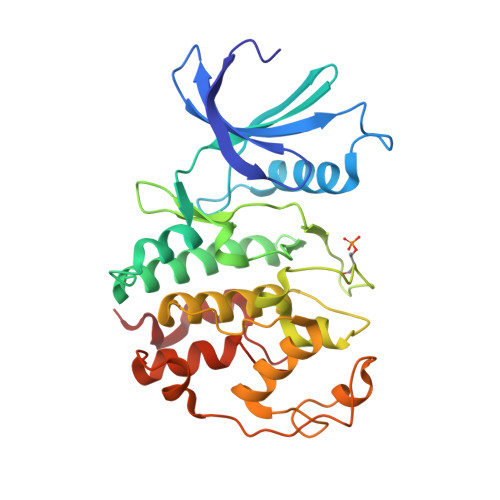
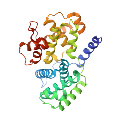
Crystal structure of phospho-CDK2 in complex with Cyclin B


Experimental Data Snapshot

  • Method: X-RAY DIFFRACTION
  • Resolution: 2.90 Å
  • R-Value Free: 0.275 
  • R-Value Work: 0.205 
  • R-Value Observed: 0.208 

Starting Model: experimental
View more details

wwPDB Validation   3D Report Full Report


This is version 1.4 of the entry. See complete history


Literature

Cyclin B and cyclin A confer different substrate recognition properties on CDK2.

Brown, N.R.Lowe, E.D.Petri, E.Skamnaki, V.Antrobus, R.Johnson, L.N.

(2007) Cell Cycle 6: 1350-1359

  • DOI: https://doi.org/10.4161/cc.6.11.4278
  • Primary Citation of Related Structures:  
    2JGZ

  • PubMed Abstract: 

    The transitions of the cell cycle are regulated by the cyclin dependent protein kinases (CDKs). The cyclins activate their respective CDKs and confer substrate recognition properties. We report the structure of phospho-CDK2/cyclin B and show that cyclin B confers M phase-like properties on CDK2, the kinase that is usually associated with S phase. Cyclin B produces an almost identical activated conformation of CDK2 as that produced by cyclin A. There are differences between cyclin A and cyclin B at the recruitment site, which in cyclin A is used to recruit substrates containing an RXL motif. Because of sequence differences this site in cyclin B binds RXL motifs more weakly than in cyclin A. Despite similarity in kinase structures, phospho-CDK2/cyclin B phosphorylates substrates, such as nuclear lamin and a model peptide derived from p107, at sequences SPXX that differ from the canonical CDK2/cyclin A substrate recognition motif, SPXK. CDK2/cyclin B phosphorylation at these non-canonical sites is not dependent on the presence of a RXL recruitment motif. The p107 peptide contains two SP motifs each followed by a non-canonical sequence of which only one site (Ser640) is phosphorylated by pCDK2/cyclin A while two sites are phosphorylated by pCDK2/cyclin B. The second site is too close to the RXL motif to allow the cyclin A recruitment site to be effective, as previous work has shown that there must be at least 16 residues between the catalytic site serine and the RXL motif. Thus the cyclins A and B in addition to their role in promoting the activatory conformational switch in CDK2, also provide differential substrate specificity.


  • Organizational Affiliation

    Laboratory of Molecular Biophysics, Department of Biochemistry, University of Oxford, Oxford, UK.


Macromolecules
Find similar proteins by:  (by identity cutoff)  |  3D Structure
Entity ID: 1
MoleculeChains Sequence LengthOrganismDetailsImage
CELL DIVISION PROTEIN KINASE 2289Homo sapiensMutation(s): 0 
EC: 2.7.1
UniProt & NIH Common Fund Data Resources
Find proteins for P24941 (Homo sapiens)
Explore P24941 
Go to UniProtKB:  P24941
PHAROS:  P24941
GTEx:  ENSG00000123374 
Entity Groups  
Sequence Clusters30% Identity50% Identity70% Identity90% Identity95% Identity100% Identity
UniProt GroupP24941
Sequence Annotations
Expand
  • Reference Sequence
Find similar proteins by:  (by identity cutoff)  |  3D Structure
Entity ID: 2
MoleculeChains Sequence LengthOrganismDetailsImage
G2/MITOTIC-SPECIFIC CYCLIN-B1260Homo sapiensMutation(s): 0 
UniProt & NIH Common Fund Data Resources
Find proteins for P14635 (Homo sapiens)
Explore P14635 
Go to UniProtKB:  P14635
PHAROS:  P14635
GTEx:  ENSG00000134057 
Entity Groups  
Sequence Clusters30% Identity50% Identity70% Identity90% Identity95% Identity100% Identity
UniProt GroupP14635
Sequence Annotations
Expand
  • Reference Sequence
Small Molecules
Modified Residues  1 Unique
IDChains TypeFormula2D DiagramParent
TPO
Query on TPO
A
L-PEPTIDE LINKINGC4 H10 N O6 PTHR
Experimental Data & Validation

Experimental Data

  • Method: X-RAY DIFFRACTION
  • Resolution: 2.90 Å
  • R-Value Free: 0.275 
  • R-Value Work: 0.205 
  • R-Value Observed: 0.208 
  • Space Group: P 61 2 2
Unit Cell:
Length ( Å )Angle ( ˚ )
a = 104.78α = 90
b = 104.78β = 90
c = 251.635γ = 120
Software Package:
Software NamePurpose
REFMACrefinement
MOSFLMdata reduction
SCALAdata scaling
PHASERphasing

Structure Validation

View Full Validation Report


Entry History 

Deposition Data

Revision History  (Full details and data files)

  • Version 1.0: 2007-05-22
    Type: Initial release
  • Version 1.1: 2011-05-08
    Changes: Version format compliance
  • Version 1.2: 2011-07-13
    Changes: Version format compliance
  • Version 1.3: 2019-10-09
    Changes: Data collection, Database references, Derived calculations, Other
  • Version 1.4: 2023-12-13
    Changes: Data collection, Database references, Refinement description